Otitis media
Otitis media. Terminology. Otitis Media: inflammation of the middle ear cleft or mucosa. Acute Less than 6 weeks Chronic More than 6 weeks Recurrent acute otitis media 3 episodes/6 months or 4 or more episodes/1 year


Otitis media
E N D
Presentation Transcript
Terminology Otitis Media: inflammation of the middle ear cleft or mucosa. Acute Less than 6 weeks Chronic More than 6 weeks Recurrent acute otitis media 3 episodes/6 months or 4 or more episodes/1 year Otitis media with effusion: fluid in the middle ear without signs or symptoms of infection.
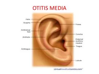
Middle ear cleft contains: Middle ear cavity Eustachian tube Mastoid antrum Mastoid air cells Aditus Atic
Otitis media with effusion (OME) • Accumulation of fluid in the middle ear • Following an episode of Otitis media • It is not necessary to have a prior episode of acute OM. • Middle ear effusion short-lived & resolves completely no need for treatment. • OME / glue ear fluid persists with an intact ear drum (no perforation) 3 months or more. • Affects most children at one time or another in up to 1/3 • Persist for 3 months or more • Commoner in winter & small children • Cause significant deafness if left untreated • May result in permanent middle-ear changes.
Etiology AOM (most imp) Adenoid Allergic rhinitis Cleft palate Down syndrome Passive smoker Early exposure to pathogens Pt’s with mucociliary disorder
Symptoms: Conductive deafness Discomfort but not pain Sometimes tinnitus. Signs: Otoscope: Dull yellow fluid behind the ear drum Audiogram: flat curve
Management • Improve spontaneously • Treat predisposing condition (allergic rhinitis or cleft palate) • Myringostomy& grommet tube • Puncture of the drum • Aspiration of the fluid • Insertion of a small tube (grommet) in the eardrum done under general anesthesia.
OME in adults • My follow • URTI • Sudden change in pressure (deep sea diving or a rapid descent from an aircraft). • Improvement is spontaneous & gradual my take up to 6wks. • Rarely a presentation of nasopharyngeal malignancy.
Chronic otitis media Inflammation of the middle ear To be called chronic it needs to last for more than 6 weeks. Usually preceded by an acute infection either acute ottits media or a viral URTI The most common age for chronic ottits media is between the ages of 3-6 years old
Causes & predisposing factors Late onset or inappropriate antibiotic treatment of AOM URTI Allergic rhinitis Sinusitis Adenoid hypertrophy Lowered Resistance (malnutrition & anemia) Short period breastfeeding Long time group child care Eustachian tube deformity Nasal septum deviation Cleft palate
Symptoms Conductive deafness Vertigo Tinnitus Ear discharge (which maybe foul smelling when there is a cholestoma present) Etiologies P. aerugenosa Proteus E.coli H. influenza
Serous OME The most common cause of serous OME is children is an enlarged adenoid Stages: URTI or acute otitis media that leads to Fluid collection in middle ear & obstruction of eutachian tube this leads to tympanic membrane retraction. Fluid become pus and glue like and that leads to conductive hearing impairment & pain which eventually will end with necrosis and tympanic membrane perforation. Could end up with mastoiditis (if untreated) Management Systemic decongestants Nasal drops Myringotomy (if the above 2 failed), tiny incision done in the ear drum to relief pressure and drain pus.
Tubo-tympanicotitis media(Safe type without cholestoma) • Acute otitis media which leads to a permanent perforation that causes muco-purulent discharge. • Infection is limited to the mucosa (ant. Inf.) • With this type there is no risk of bone destruction • The perforation of the membrane is mostly directed centerally Management • Clean the ear by syringing or hydrogen peroxide. • Local antibiotic (when the ear is totally clean and dry) • Surgery (if medical treatment failed) • Myringoplasty: repair of tympanic membrane perforation & ossicles are intact (most used graft is autologoustemporalis fascia) • Tympanoplasty: repair of tympanic membrane & ossicles.
Atico-antral chronic otitis media(With cholestoma) Life threatening (intra & extra cranial complications) spreads by bone destruction (mastoid, tympanic ring, ossicles ) Perforation is posterio-superior Discharge is usually persistent and often foul smelling. There is granulation due to osteitis. Aural polyps formed by granulation tissues Associated with chlesteatoma: Management: Regular aural toilet in early cases of annular osteitis may be adequate to prevent progression. Surgical removal of cholestetoma Treatment of complications
Central pars tensa tympanic membrane perforation with a healthy middle ear membrane.
How to differentiate between tubo-typmanic and attico-antral clinically The first difference can be observed in discharge The second difference can be observed in the position of the perforation itself The third difference can be observed with the presence of a cholestatmoa The fourth difference can be observed with severity of deafness
Cholesteatoma Epithelial cells collection in the middle ear cleft basically an epidermoidcyst. Produces mass effect on the structures that are present in the cleft Treatment is by surgical excison Why does bone erosion happen in chronic otitis media? Pressure theory Enzymatic theory (acid phosphatase, collagenase &other proteolytic enzymes) which are present in the cholestatoma Pyogenicosteitis (Pyogneic bacteria may release enzymes)