Download
1 / 34
420 likes | 811 Views
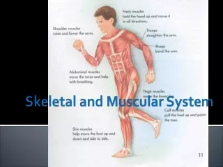
Muscular and Skeletal System. Powerpoint #2 Unit 8 – Chapters 35/36 Working together to create movement . Skeletal System. Structures : Bones Cartilage Ligaments Tendons. Skeletal System. Function: Supports body Protects internal organs Allows for movement

E N D
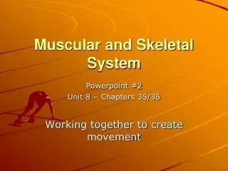
Muscular and Skeletal System Powerpoint #2 Unit 8 – Chapters 35/36 Working together to create movement
Skeletal System Structures: • Bones • Cartilage • Ligaments • Tendons
Skeletal System Function: • Supports body • Protects internal organs • Allows for movement • Stores mineral reserves • Provides a site for blood cell formation
How many bones in a human Skeleton? • 206 • How many in babies? ~ 300 • What are bones? • A solid network of living cells and protein fibers that are surrounded by deposits of calcium salts
Bones: 3 Parts • Spongy bone • Not soft or spongy • Very strong • Structure resembles the supporting structure of bridges. • Strong but lightweight
Compact bone: • Very dense (no spaces like spongy bone) • Outer portion of bone • Contains Haversian canal for veins and arteries to run through
Bone Marrow: • Soft tissue • Found in bone cavities • Yellow Marrow: fat cells • Red marrow: • makes red blood cells, platelets and most white blood cells • ~1/2 red marrow turns into yellow in adults • If severe blood loss, some yellow can turn back to red
Development of bone: • Cartilage: Tough, elastic, connective tissue • Found in: ears, between bones, larynx, and other various places.
Development of Bone • http://www.youtube.com/watch?v=78RBpWSOl08 • Ossification: cartilage replaced by bone • Osteoblasts:cells that build bone • Osteoclasts:cells that break down bone • growth Plates: found in most long bones (leg and arm bones) cartilage continues to grow here until completely replaced by bone during adolescence
Types of Joints • Ball and socket
Types of Joints • Hinge
Types of Joints • Saddle
Types of Joints • Pivot
Structure of Joints • Ligaments: Connect Bone to Bone
Structure of Joints • Bursa:
Skeletal muscle Cardiac muscle Smooth muscle Types of Muscle • The human body is comprised of 324 muscles • Muscle makes up 30-35% (in women) and 42-47% (in men) of body mass. Three types of muscle:
A. Skeletal (Striated) Muscle • Connects the various parts of the skeleton through one or more connective tissue tendons • During muscle contraction, skeletal muscle shortens and moves various parts of the skeleton • Activated through signals carried to the muscles via nerves voluntary control • Repeated activation of a skeletal muscle can lead to fatigue • Can have many nuclei
Skeletal Muscles work in PAIRSBending or straightening of elbow requires the coordinated interplay of the biceps and triceps muscles
B. Smooth Muscle • Located in the blood vessels, the respiratory tract, the iris of the eye, the gastro-intestinal tract • The contractions are slow and uniform • Is fatigue resistant • Activation is involuntary • Has one nucleus
C. Cardiac Muscle • Has characteristics of both skeletal and smooth muscle • Functions to provide the contractile activity of the heart • Is very fatigue resistant • Activation of cardiac muscle is involuntary (like smooth muscle) • Can have 2 nuclei, usually has 1
Components of skeletal muscle myofibril muscle fiber muscle fiber bundle
Muscle Fibers • Cylinder-shaped cells that make up skeletal muscle • Each myofibril is made up of a number of myofilaments • Diameterof fiber(0.05-0.10 mm) Lengthof fiber(appr. 15 cm) • Each fibercontains contractile machinery and cell organelles • Group of fiberactivated via same nerve: motor unit • Each fiberhas capillaries that supply nutrients and eliminate waste • Divided into functional units called sarcomeres
High microscope magnification of sarcomereswithin a myofibril
Muscle Contraction • Organized in series (attached end to end) • Two types of protein myofilaments: - Actin:thin filament - Myosin: thick filament • Projecting from each myosin are tiny contractile myosin bridges
Muscle Contraction • During muscle contraction the myofilaments myosin and actin slide toward each other and overlap. This shortens the sacromere and the entire muscle. Muscle cells are "shocked" by nerve impulses from motor neurons.
(a) At rest Muscle Contraction The filaments slide together because myosin attaches to actinand pulls on it. Myosin head (H) attaches to actin filament (A), forming a cross bridge. After the cross bridge is formed the myosin head bends, pulling on the actin filaments and causing them to slide: Muscle contraction is a little like climbing a rope. The cross bridge cycle is: grab -> pull -> release, repeated over and over b) Contraction
Tendons • Connect Muscle to Bone