Hematopathology Case 2
40 likes | 52 Views
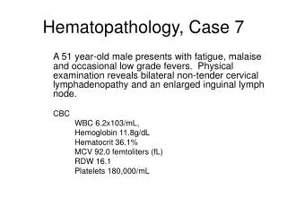
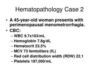
This case features a 45-year-old woman with perimenopausal menometrorrhagia. CBC results show low hemoglobin, hematocrit, and MCV with increased RDW, indicating iron deficiency anemia. Peripheral blood smear reveals hypochromic and microcytic red blood cells. This comparison to a normal smear highlights the abnormalities in iron deficiency anemia.

Hematopathology Case 2
E N D
Presentation Transcript
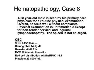
Hematopathology Case 2 • A 45-year-old woman presents with perimenopausal menometrorrhagia. • CBC: • WBC 9.7x10³/mL • Hemoglobin 7.8g/dL • Hematocrit 23.5% • MCV 73 femtoliters (fL) • Red cell distribution width (RDW) 22.1 • Platelets 187,000/mL
Neutrophil Most of the red blood cells are hypochromic (increased central pallor). Many of the red blood cells are microcytic (arrows). History, CBC, peripheral smear are consistent with iron deficiency anemia
Iron deficiency anemia Compare iron defeciency anemia on left to a normal smear on right.