Download
1 / 51
510 likes | 613 Views
Explore the intricate workings of neurons and nerve impulses in the nervous system. Learn about different types of neurons, synaptic transmission, and the functions of the central and peripheral nervous systems.

E N D
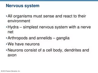
Nervous system • All organisms must sense and react to their environment • Hydra – simplest nervous system with a nerve net • Arthropods and annelids – ganglia • We have neurons • Neurons consist of a cell body, dendrites and axon
Types of neurons • Sensory – receive impulses from the environment and bring them to the body • Motor – transmits the impulse to the muscles or glands in response • Interneurons – link sensory and motor neurons – they are found in the brain and spinal cord
Cell body Dendrite Mitochondrion Nissl substance Axon hillock Axon Collateral branch Neurofibrils Nucleus One Schwann cell Node of Ranvier Axon terminal Schwann cells, forming the myelin sheath on axon (a) Figure 7.4a
Reflex arc Central process (axon) Sensory neuron Spinal cord (central nervous system) Cell body Ganglion Dendrites Peripheral process (axon) Afferent transmission Interneuron (association neuron) Peripheral nervous system Receptors Efferent transmission Motor neuron To effectors (muscles and glands) Figure 7.6
Nerve Impulses • Resting neuron • The plasma membrane at rest is polarized • Fewer positive ions are inside the cell than outside the cell
[Na+] + + + + + + + 1 + Resting membrane is polarized. In the resting state, the external face of the membrane is slightly positive; its internal face is slightly negative. The chief extracellular ion is sodium (Na+), whereas the chief intracellular ion is potassium (K+). The membrane is relatively impermeable to both ions. – – – – – – – – [K+] – – – – – – – + + + + + + + Figure 7.9, step 1
Na+ + + + + + + + 2 Stimulus initiates local depolarization. A stimulus changes the permeability of a local "patch" of the membrane, and sodium ions diffuse rapidly into the cell. This changes the polarity of the membrane (the inside becomes more positive; the outside becomes more negative) at that site. + – – – – – – – – + – – – – – – – + + + + + + + Figure 7.9, step 2
Na+ + + – + + – + 3 + Depolarization and generation of an action potential. If the stimulus is strong enough, depolarization causes membrane polarity to be completely reversed and an action potential is initiated. – – – – – + + – + – – – – – + + – + + – + + + Figure 7.9, step 3
– – – + – – + + 4 Propagation of the action potential. Depolarization of the first membrane patch causes permeability changes in the adjacent membrane, and the events described in step are repeated. Thus, the action potential propagates rapidly along the entire length of the membrane. + – + – + – + + 2 – + – + + – + – – – + – + + Figure 7.9, step 4
+ – + – + – + 5 + – Repolarization. Potassium ions diffuse out of the cell as the membrane permeability changes again, restoring the negative charge on the inside of the membrane and the positive charge on the outside surface. Repolarization occurs in the same direction as depolarization. – + + – + – – + K+ – + – + – + + – + – + – + – Figure 7.9, step 5
Cell exterior Na+ Na+ Na+ – K+ pump Na+ 6 Initial ionic conditions restored. The ionic conditions of the resting state are restored later by the activity of the sodium-potassium pump. Three sodium ions are ejected for every two potassium ions carried back into the cell. K+ Na+Diffusion Plasma membrane K+Diffusion K+ K+ Cell interior K+ Figure 7.9, step 6
Parts of the Nervous System Central Nervous System • Brain and spinal cord Peripheral Nervous System • All other nerves • Somatic and autonomic • Somatic – voluntary activities – skeletal muscle contraction • Autonomic – involuntary activities – digestion/heartbeat • These systems sometimes work together
Nervous Tissue: Neurons Neurons = nerve cells Cells specialized to transmit messages Major regions of neurons Cell body—nucleus and metabolic center of the cell Processes—fibers that extend from the cell body
Nervous Tissue: Neurons Cell body Nissl bodies Specialized rough endoplasmic reticulum Neurofibrils Intermediate cytoskeleton Maintains cell shape Nucleus with large nucleolus
Cell body Dendrite Mitochondrion Nissl substance Axon hillock Axon Collateral branch Neurofibrils Nucleus One Schwann cell Node of Ranvier Axon terminal Schwann cells, forming the myelin sheath on axon (a) Figure 7.4a
Nervous Tissue: Neurons Processes outside the cell body Dendrites—conduct impulses toward the cell body Neurons may have hundreds of dendrites Axons—conduct impulses away from the cell body Neurons have only one axon arising from the cell body at the axon hillock
Nervous Tissue: Neurons Axons End in axon terminals Axon terminals contain vesicles with neurotransmitters Axon terminals are separated from the next neuron by a gap Synaptic cleft—gap between adjacent neurons Synapse—junction between nerves
Nervous Tissue: Neurons Myelin sheath—whitish, fatty material covering axons Schwann cells—produce myelin sheaths in around axons (PNS) Nodes of Ranvier—gaps in myelin sheath along the axon – saltatory conduction Oligodendrocytes—produce myelin sheaths around axons of the CNS
Transmission of a Signal at Synapses • When the action potential reaches the axon terminal, the electrical charge opens calcium channels
Axon of transmitting neuron Receiving neuron Action potential arrives. 1 Dendrite Vesicles Axon terminal Synaptic cleft Figure 7.10, step 1
Transmitting neuron 2 Vesicle fuses with plasma membrane. Synaptic cleft Ion channels Neurotransmitter molecules Receiving neuron Figure 7.10, step 2
Transmitting neuron 2 Vesicle fuses with plasma membrane. 3 Neurotrans- mitter is released into synaptic cleft. Synaptic cleft Ion channels Neurotransmitter molecules Receiving neuron Figure 7.10, step 3
Transmitting neuron 2 Vesicle fuses with plasma membrane. 4 Neurotrans- mitter binds to receptor on receiving neuron's membrane. 3 Neurotrans- mitter is released into synaptic cleft. Synaptic cleft Ion channels Neurotransmitter molecules Receiving neuron Figure 7.10, step 4
Transmission of a Signal at Synapses If enough neurotransmitter is released, graded potential will be generated Inhibitory or excitatory Eventually an action potential (nerve impulse) will occur in the neuron beyond the synapse
Neurotransmitter Receptor Na+ 5 Ion channel opens. Figure 7.10, step 5
Neurotransmitter is broken down and released. Na+ 6 Ion channel closes. Figure 7.10, step 6
Transmission of a Signal at Synapses The electrical changes prompted by neurotransmitter binding are brief The neurotransmitter is quickly removed from the synapse
Acetylcholine • Neurotransmitter that is released from the axon as a result of Ca2+ entering the axon terminal • Stimulates muscle contraction • Active in parasympathetic nervous system • Norepinephrine and GABA are other neurotransmitters
Central nervous system Peripheral nervous system Effector organs Acetylcholine Skeletal muscle Somatic nervous system Smooth muscle (e.g., in stomach) Norepinephrine Acetylcholine Ganglion Sympathetic division Epinephrine and norepinephrine Acetylcholine Autonomic nervous system Blood vessel Glands Adrenal medulla Acetylcholine Cardiac muscle Parasympathetic division Ganglion KEY: Postganglionic axons (sympathetic) Preganglionic axons (parasympathetic) Postganglionic axons (parasympathetic) Preganglionic axons (sympathetic) Myelination Figure 7.27
Axon terminals at neuromuscular junctions Spinal cord Motor unit 2 Motor unit 1 Nerve Axon ofmotorneuron Motor neuroncell bodies Muscle Muscle fibers (a) Figure 6.4a
Synaptic vesicle containing ACh Action potential reaches axonterminal of motor neuron. 1 Axon terminal of motor neuron Mitochondrion Ca2+ 2 Calcium (Ca2+) channelsopen and Ca2+ enters the axon terminal. Ca2+ Synapticcleft Sarcolemma Fusing synapticvesicle Sarcoplasmof muscle fiber 3 Ca2+ entry causes somesynaptic vesicles to release theircontents (acetylcholine, a neurotransmitter) by exocytosis. ACh Folds ofsarcolemma AChreceptor 4 Acetylcholine diffuses acrossthe synaptic cleft and binds toreceptors in the sarcolemma. Figure 6.5, step 4
Ion channel insarcolemma opens;ions pass. K+ Na+ 5 ACh binds and channels open that allow simultaneous passage of Na+ into the muscle fiber and K+ out of the muscle fiber. More Na+ ions enter than K+ ions leave and this produces a local change in the electrical conditions of the membrane (depolarization), which eventually leads to an action potential. Figure 6.5, step 5
ACh Degraded ACh Ion channel closed;ions cannot pass. Na+ ACh effects are ended by itsbreakdown in the synaptic cleft bythe enzyme acetylcholinesterase. 6 Acetylcholinesterase K+ Figure 6.5, step 6
Short term More prolonged Stress Hypothalamus Releasing hormones Nerve impulses Spinal cord Corticotropic cells of anterior pituitary ACTH Adrenal cortex Preganglionic sympathetic fibers Adrenal medulla Mineralocorticoids Glucocorticoids Long-term stress response Short-term stress response 1. Increased heart rate 2. Increased blood pressure 3. Liver converts glycogen to glucose and releases glucose to blood 4. Dilation of bronchioles 5. Changes in blood flow patterns, leading to increased alertness and decreased digestive and kidney activity 6. Increased metabolic rate 1. Proteins and fats converted to glucose or broken down for energy 2. Increased blood sugar 3. Suppression of immune system Catecholamines (epinephrine and norepinephrine) 1. Retention of sodium and water by kidneys 2. Increased blood volume and blood pressure Figure 9.13
Brain • Cerebrum – convoluted folds, two halves • Corpus callosum – allows the cerebral hemispheres to communicate • Left side – dominant in math, language, logic • Right – nonverbal thinking and image recognition • Hypothalamus and pituitary gland – hormone release, limbic system (thirst, pain, sexual desire) • Cerebellum – coordination and muscle movement
Skeletal Muscle Functions Produce movement Maintain posture Stabilize joints Generate heat
Microscopic Anatomy of Skeletal Muscle Sarcolemma—specialized plasma membrane Myofibrils—long organelles inside muscle cell Sarcoplasmic reticulum—specialized smooth endoplasmic reticulum
Sarcolemma Myofibril Nucleus Light(I) band Dark(A) band (a) Segment of a muscle fiber (cell) Figure 6.3a
Z disc H zone Z disc Thin (actin) filament Thick (myosin) filament (b) Myofibril or fibril (complex organelle composed of bundles of myofilaments) I band M line I band A band Figure 6.3b
Microscopic Anatomy of Skeletal Muscle Sarcomere—contractile unit of a muscle fiber Organization of the sarcomere Myofilaments Thick filaments = myosin filaments Thin filaments = actin filaments
Sarcomere M line Z disc Z disc Thin (actin) filament Thick (myosin) filament (c) Sarcomere (segment of a myofibril) Figure 6.3c
The Nerve Stimulus and Action Potential Skeletal muscles must be stimulated by a motor neuron (nerve cell) to contract Motor unit—one motor neuron and all the skeletal muscle cells stimulated by that neuron
Axon terminals at neuromuscular junctions Spinal cord Motor unit 2 Motor unit 1 Nerve Axon ofmotorneuron Motor neuroncell bodies Muscle Muscle fibers (a) Figure 6.4a
The Sliding Filament Theory of Muscle Contraction Activation by nerve causes myosin heads (cross bridges) to attach to binding sites on the thin filament Myosin heads then bind to the next site of the thin filament and pull them toward the center of the sarcomere This continued action causes a sliding of the myosin along the actin The result is that the muscle is shortened (contracted)
Actin Myosin Z Z H A I I (a) Z Z I A I (b) Figure 6.7a–b
Protein complex In a relaxed muscle cell, the regulatory proteins formingpart of the actin myofilaments prevent myosin binding(see a). When an action potential (AP) sweeps along itssarcolemma and a muscle cell is excited, calcium ions(Ca2+) are released from intracellular storage areas (thesacs of the sarcoplasmic reticulum). Myosinmyofilament Actinmyofilament (a) Figure 6.8a
Myosin-binding site The flood of calcium acts as the final trigger forcontraction, because as calcium binds to the regulatoryproteins on the actin filaments, the proteins undergo a change in both their shape and their position on the thinfilaments. This action exposes myosin-binding sites onthe actin, to which the myosin heads can attach (see b),and the myosin heads immediately begin seeking out binding sites. Ca2+ Upper part of thick filament only (b) Figure 6.8b


