Download
1 / 25
300 likes | 1.01k Views
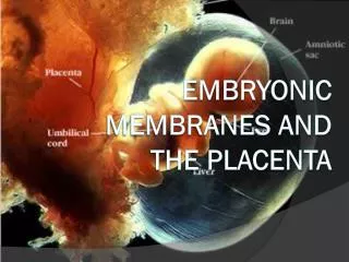
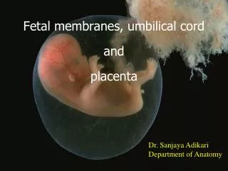
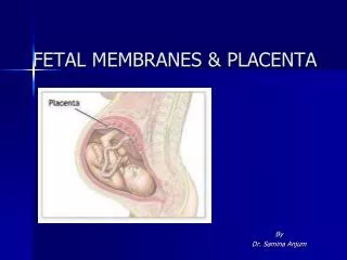
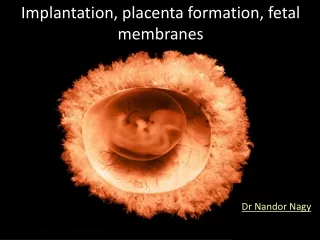
FETAL MEMBRANES AND PLACENTA BY. PROF MASOOD AHMED MBBS, MPHIL PHD. Relative Size of Human Conceptus. Implantation of the Blastocyst. Implantation of the Blastocyst. FETAL MEMBRANES. REFRESH FEW TERMS DECIDUA BASALIS, CAPULARIS AND PARIETALIS VILLI FORMATION

E N D
FETAL MEMBRANES AND PLACENTABY PROF MASOOD AHMED MBBS, MPHIL PHD
FETAL MEMBRANES REFRESH FEW TERMS DECIDUA BASALIS, CAPULARIS AND PARIETALIS VILLI FORMATION CYTOTRPHOBLASTIC SHELL FORMATION CHORIONIC MEMBRANE PARTS CHORION FURONDOSUM AND CHORION LAEVE AMNIOCHORIONIC MEMBRANE
Embryonic Membranes • Amnion – Epiblast cells form a transparent membrane filled with amniotic fluid • Provides a buoyant environment that protects the embryo • Helps maintain a constant homeostatic temperature • Amniotic fluid comes from maternal blood, and later, fetal urine
CYTOTROPHOBLASTIC SHELL FORMATIONPLACENTAL BARRIER/ MEMBRANE
Embryonic Membranes • Allantois – a small outpocketing at the caudal end of the yolk sac • Structural base for the umbilical cord • Becomes part of the urinary bladder • Chorion – helps form the placenta • Encloses the embryonic body and all other membranes
Placentation • The chorion develops fingerlike villi, which: • Become vascularized • Extend to the embryo as umbilical arteries and veins • Lie immersed in maternal blood • Deciduabasalis – part of the endometrium that lies between the chorionic villi and the stratum basalis
UTERO PLACENTAL CIRCULATION –12TH DAY CIRCULATION • 4TH MONTH CHORION FRONDOSUM AND DECIDUA BASALIS • 5TH MONTH DECIDUAL SEPTA AND COTYLEDON FORMATION CONTIOUS FLOW IN INTERVILLOUS SPACE
PLACENTAL SURFACES • FETAL SURFACE • CHORIONIC VESSELLS CONVERGING TOWARDS UMBLICAL CORD • 2-ARTERIES AND ONE VEIN • CHORION COVERED BY AMNION • ECCENTRIC ATTACHMENT OF UMBILICAL CORD • MATERNAL SURFACE • 15-20 COTYLEDONS • Functional type • hemochorial
FULL TERM PLACENTA • 15-20% INNER SURFACE COVERED WITH PLACENTA • ALL THE TIME SYNCITIAL SURFACE SEPARATES THE MATERNAL BLOOD • COTYLEDONS- CORE OF ENDOMETRIAL TISSUE COVERED BY SYNSITIUM • 15-20 IN NUMBER • SHAPE-DISCOID • SURFACE AREA 15-20cm • THICKNESS-3cm • WEIGHT-500-600gms
Placenta • 80-100 MATERNAL ENDOMETRIALSPIRAL ARTERIES SUPPLYING VILLI • A TOTAL 150 ML BLOOD IN PLACENTAL CIRCULATION • REPL;ENISHES 3-4 TIMES / MINUTE • PLACENTAL BARRIER • SEPARATES MATERNAL AND FETAL BLOOD • COMPRISED OF • ENDOTHELIAL LINING OF VILLI CAPPILARIES • C.T IN VILLI • TROPHOBLASTIC LAYER • CYTO TROPHOBLAST LAYER • 4TH MONTHS ONWARD • INCREASED EXCHANGE BY • C.T AND TROPHOBLAST LAYER DISAPPEAR • CLOSE TO END OF PREGNANCY • DECREASE EXCHANGE BY • INCREASE FIBROUS TISSUE IN VILLI • FIBRINOID DEPOSIT ON SURFACE OF VILLI • OBLITERATION OF SMALL CAPPILARIES
Variations of placenta • Normally---hemochorial and eccenteric • Variations • velamentous –when umblical vessels divide before and branches come to placenta • Marginal- when umblical vessels join placenta at margin • Succenturiate- when additional small plcental lobe is present or separate small placenta is present • accreta- when placental reaches up to basal layer of endometrium • Increta- when placental infiltrates in the myometrium • Percreta- when placenta infiltrates up to connective tissue and crosses the muscule layer
AMNIOTIC FLUID • SYNTHESIZED BY • CLEAR WATERY FLUID • MAINLY BY • MATERNAL CIRCULATION • PARTLY BY AMNIOCYTES • 30-ML—10TH WEEK • 350 ML—20TH WEEK • 800-1000 ML---37TH WEEK • REPLACED /3-4 HRS • 5TH MONTH ONWARD • SWALLOWING • AND ADDITION OF URIN • FUNCTIONS • ABSORBS JOLTS • ALLOW FETAL MOVEMENTS • HORMONE PRODUCTION • AVOIDS ADHERENCE • Polyhydroamnios • Amount of amniotic fluid • more than 1500 ml eg • Esophageal atresi, anencephaly • Oligohydroamnios • Amount of amniotic fluid • less than 400 ml eg • Renal agenesis
Membranes in twins • DIZYGOTIC OR FRATERNAL twins • Separate amniotic and chorionic membranes and placenta • May be different sex, blood groups, features
Monozygotic twins • If twins form by the division of zygot at two cell stage with the formation of two separate blastocyst • Two Separate amniotic and chorionic membranes and placenta • Features,Sex and blood groups same
If twins form by the division of inner cell mass in blastocyst • Two Separate amniotic cavities • Common chorionic membranes and placenta • Features, Sex and blood groups same
If twins form by the late division of inner cell mass in blastocyst or by division of bilaminar germ disc • Common amniotic, chorionic membranes and placenta • Features, Sex and blood groups same