Download
1 / 17
220 likes | 1.07k Views
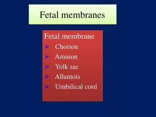
Fetal membranes. Fetal membrane Chorion Amnion Yolk sac Allantois Umbilical cord.

E N D
Fetal membranes Fetal membrane • Chorion • Amnion • Yolk sac • Allantois • Umbilical cord
Yolk sac:it is a membranous sac attached to the embryo, convey nourishment to the embryo (e.g. in birds), in humans, Incorporate into the endoderm of embryo as a primordial gut and the primordial germ cells appear in the endodermal lining of the wall of the yolk sac in the 3rd week
It is large at 32 days (at early development) • by 10th week, regresses to 0.5 cm as a remnant structure which is connected to the midgut by a narrow yolk stalk • at 20 weeks becomes very small, usually not visible thereafter.
Abnormalities: Sometimes it persists throughout the pregnancy but of no significance In about 2% of adults the proximal intra-abdominal part of yolk stalk persists as an ileal diverticulum or Meckel diverticulum ①Meckel’s diverticulum ② Umbilical fistula
allantois, an extra-embryonic membrane of reptiles, birds, and mammals arising as a pouch, or sac, from the hindgut In the 3rd week it appears as a tubular diverticulum from the caudal wall of yolk sac that extends into the connecting stalk During the 2nd month, the extraembryonic part of the allantois degenerates
Functions of Allantois Blood formation occurs in the wall during the 3rd to 5th week Its blood vessels persist as the umbilical vein and arteries BecomesUrachusand after birth is transformed into median umbilical ligament extends from the apex of the bladder to the umbilicus
Abnormality: Urachal fistula
Amniotic Fluid The amniotic cavity is filled with a clear, watery fluid is produced in part by amniotic cells but is derived primarily from maternal blood The amount of fluid increases from approximately 30 ml at 10 weeks of gestation to 450 ml at 20 weeks to 800 to 1000 ml at 37 weeks serves as a protective cushion. The fluid 1- prevents adherence of the embryo to the amnion 2-allows for fetal movements The volume of amniotic fluid is replaced every 3 hours From the beginning of the fifth month, the fetus swallows its own amniotic fluid and it is estimated that it drinks about 400 ml a day, about half of the total amount Fetal urine is added daily to the amniotic fluid in the fifth month but this urine is mostly water, since the placenta is functioning as an exchange for metabolic wastes
During childbirth, the amnio-chorionic membrane forms a hydrostatic wedge that helps to dilate the cervical canal.
1-Hydramnios or polyhydramnios is the term used to describe an excess of amniotic fluid (1500–2000 ml) Primary causes of hydramnios include idiopathic causes (35%) maternal diabetes (25%) congenital malformations including central nervous system disorders (e.g., anencephaly) and Gastrointestinal defects (atresias, e.g., esophageal) that prevent the infant from swallowing the fluid 2-Oligohydramnios refers to a decreased amount (less than 400 ml) Oligohydramnios is a rare occurrence that may result from renal agenesis Premature rupture of the amnion, the most common cause of preterm labor occurs in 10% of pregnancies
With growth of the chorionic vesicle the decidua capsularis becomes stretched and degenerates Subsequently the chorion laeve comes into contact with the uterine wall (decidua parietalis) on the opposite side of the uterus and the two fuse obliterating the uterine lumen. Similarly, fusion of the amnion and chorion to form the amniochorionic membrane obliterates the chorionic cavity It is this membrane that ruptures during labor (breaking of the water).
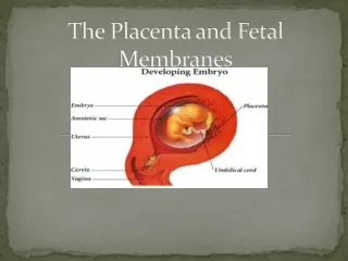
At the fifth week of development, the following structures pass through the primitive umbilical ring (1) The connecting stalk, containing the allantois and the umbilical vessels, consisting of two arteries and one vein (2) The yolk stalk (vitelline duct), accompanied by the vitelline vessels (3) The canal connecting the intraembryonic and extraembryonic cavities
During further development, the amniotic cavity enlarges rapidly at the expense of the chorionic cavity As a result the amnion begins to envelop the connecting and yolk sac stalks, crowding them together and giving rise to the primitive umbilical cord
The abdominal cavity is temporarily too small for the rapidly developing intestinal loops and some of them are pushed into the extraembryonic space in the umbilical cord. These extruding intestinal loops form a physiological umbilical hernia At approximately the end of the third month the loops are withdrawn into the body of the embryo and the cavity in the cord is obliterated. When the allantois and the vitelline duct and its vessels are also obliterated, all that remains in the cord are the umbilical vessels surrounded by the jelly of Wharton.
Summary of the Umbilical cord: • Covered with amniotic membrane • Contains 1umbilical vein 2umbilical arteries degenerated yolk sac and allantois • connects fetus with placenta • Length 50 cm Abnormality: >80 cm (long), An extremely long cord may encircle the neck of the fetus, usually without increased risk <35 cm (short) may cause difficulties during delivery by pulling the placenta from its attachment in the uterus
Umbilical vessels are longer than the cord, so twisting and bending of the vessels are common They frequently form loops, producing false knots, that are of no significance In about 1% of pregnancies, true knots form in the cord and cause fetal death