Download
1 / 75
760 likes | 935 Views
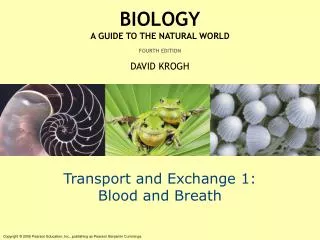
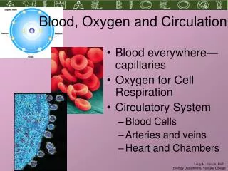
Blood and Circulation (Cardiovascular System). Blood. Functions: Transports material between body cells and external environment oxygen for CR, carbon dioxide away, nutrients, ions, hormones, immune cells and antibodies Distributes heat

E N D
Blood Functions: • Transports material between body cells and external environment • oxygen for CR, carbon dioxide away, nutrients, ions, hormones, immune cells and antibodies • Distributes heat • Protection of the body by white blood cells and antibodies that circulate in the blood and defend the body against foreign material. • Clotting mechanisms are also present that protect the body from blood loss after injuries.
Blood I. Blood composition (connective tissue – cells in a liquid matrix - ~5-6 L/person (8% of body weight) Hematocrit What would having a greater percent of RBCs mean? Where have you seen this in the news lately?
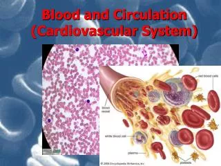
Blood Composition A. Plasma (Liquid) – 54% • H2O (92%) • Proteins • Fibrinogen - clotting • Globulins - antibodies • Albumin - osmostic balance b/w blood and tissue • Others – hormones, enzymes • Monomers: aa, glucose, fatty acids, nucleic acids, and vitamins from digested food • Salts (electrolytes or ions) - maintain pH, osmotic balance, need for muscle contraction • Cellular waste (CO2, N2) and O2
Blood Composition Continued B. Formed Elements (Cells) 1. RBC’s (erythrocytes) – 45% • Fun Facts: mainly hemoglobin (gives red color when combines with O2 vs. blue color), have no nucleus so can’t divide and no mitochondria so don’t use the O2 they carry, lasts 120 days circulating body 75,000 times) • Hematocrit – amount of blood sample made up of RBCs Shape = adaptation to carry more O2 and squeeze through narrow capillaries
Blood Composition Continued B. Formed Elements (Cells) 2. WBC’s (leukocytes) – 1% • Immune Cells - protect against infection in various ways • Phagocytize bacterial cells • Produce antibodies • transported by blood to sites of infection (immune system) Granulocytes vs. Agranulocytes
Blood Composition Continued a. Granulocytes – made in bone marrow, their names is based on when stained they look granular, non-specific, life span ~12 hrs 1. Neutrophils – phagocytes (engulfs bacteria only), 1st responders, wound healing 2. Basophils – (mast cells) release histamine which promotes inflammation and allergic reactions, dilated vessels inc. blood flow (more O2), signals for more spec macrophages to come to infected site to catch pathogens 3. Eosinophils - ?? fights parasites, mediates inflammation and allergic reaction
Blood composition Continued b. Agranulocytes made in bone marrow, their names is based on when stained they lack granules 1. Monocytes – when activated become macrophages (a wandering phagocytic cell that destroys large amounts of bacteria, damaged tissue and viruses), non-specific, life span several weeks to months
Blood composition Continued 2. Lymphocytes – made in lymph glands, spleen, and thymus; specific immunity, life span years • B cells – make antibodies (proteins that attack foreign molecules), clumps pathogens to be eaten by macrophages • T cells - Cytotoxic – “killers” – kill infected cells from bacteria and viruses, tumor cells and transplanted cells (foreign) • T cells – Helper – increase B cells and T killers
Blood composition Continued 3. Platelets (Thrombocytes) – small fragments of larger shattered cells, arise from bone marrow, life span ~ 10 days • Blood clotting - release serotonin which helps to reduce blood loss from broken vessels by contracting smooth muscles in vessel walls and thromoboplastin.
Blood Composition Continued • Relative Abundance of WBC’s: • Neutrophils (54-62 %), Lymphocytes, Monocytes, Eosinophils, Basophils (<1%) • Never Let Monkeys Eat Bananas • Leukocytes are recruited and activated by cell damage or foreign tissue • Cytokines (chemical attractors of WBC’s) are released by T helpers causing chemotaxis – cells release chemicals
Leukocytes leave the blood circulation by squeezing through the walls of the blood vessels, travel through the interstitial space and enter the site of injury. basophils lymphocytes
Blood Formation • Forms in red marrow from hematopoietic stem cells in flat bones and a little in the ends of long bones • Erythropoietin (EPO) - hormone produced by the kidney maintains homeostasis by controling RBC production • Low O2 from high altitude, loss of blood (Natural or blood dopping), lung diseases causes release of erythropoietin from kidney and liver increases RBC production better oxygen carrying capability shuts off release of erythropoietin (negative feedback loop)
RBC Proteins (Blood Types) • Over 30 types of RBC proteins (antigens or agglutinogens) • Antigen – protein that induces an immune response to make antibodies (antibody generator) • Most common – A, B, O • Rh group (determines positive blood (+) if have Rh protein or negative blood (–) if missing Rh protein on RBCs) • Antibody (agglutinin) – protein produced by the immune system in response to the presence of an antigen (Ex. Anti-A) • Agglutination – clumping
Blood Types (antigen) Immune response is to make antibodies against proteins (antigens) that are foreign to the body
Rh Antigen The Rh factor is typically called the D agglutinogen. It was originally found on the red blood cells of Rhesus monkeys (hence the “Rh” factor).
Blood Transfusions • Various proteins are embedded in the surfaces of red blood cells, these proteins are inherited. • If during a transfusion an individual receives blood containing RBCs with proteins that the individual does not carry, these proteins are recognized as foreignantigens by the immune system. • Antibodies are produced that bind to the antigens and cause agglutination(clumping) and subsequent destruction of the foreign RBCs clumping allows macrophages to engulf cells and result is death. • For example, the immune system would respond if a person with A blood type (either AA or AO) receives blood of the B or AB blood type, but not of the O type. (The O type does not carry any foreign agglutinogens.)
Hemostasis – Stoppage of Bleeding from small vessels as opposed to larger = hemorrhage • 1. BV Spasm - platelets stick to broken surfaces and produce serotonin (neurotransmitter) which contracts smooth muscle causing it to contract and narrows the vessel so blood flow is shut off to that area • 2. Platelet Plug - platelets stick to endothelium & each other only when there is a small tear in bv. • 3. Blood coagulation (blood clot) 2-6 minutes- In presence of damaged bv walls or tissue, platelets produce thromoboplastin (enzyme) which cleaves prothrombin (inactive) to thrombin (active) • Thrombin links fibrinogen proteins together (small soluble fibrous proteins) and it becomes fibrin – which is large insoluble protein fibers that act like a mesh linking WBC’s and RBC’s together • Fibrin covers over the hole in the bv and traps the RBC’s
Blood Diseases • Thrombus – abnormal blood clot due to fatty deposits/plaque build up in artery walls • Embolus – clot that has broken off – can block blood supply to organs and may cause stroke or death
Blood Diseases • Leukemia – over production of WBC’s – immature and don’t work – metatstasize to liver, spleen – use up all metabolic substrates and cause tissue destruction • Polycythemia – bone marrow cancer – make too many RBC’s RBC’s > 45%
Blood Diseases • Hemophilia – lack of a clotting factor – blood clots slowly on internal bleeds – 1 hour vs. 6-8 minutes • Anemia – deficiency of RBC’s or hemoglobin (decreased O2 carrying ability) • Sickle cell anemia – genetic point mutation in hemoglobin • Aplastic Anemia – cancer – can’t produce RBC’s • Iron Deficiency Anemia
Cardiovascular System • Function: sends oxygen rich blood and nutrients to all body cells and removes waste • Blue vs. Red? Are veins actually carrying blue blood? • Misconception. Blood in your veins has very little oxygen and is a dark red color that looks almost blue when covered by your skin. Your arteries have bright red blood because it has a lot of oxygen in it that is being carried throughout your body to be used by tissues.
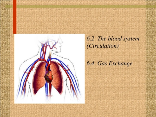
Parts of Cardiovascular System • Heart – muscular pump, ~6000 quarts/day • Vessels – organs that form a closed circuit that carries blood from heart to body cells and back again. • Arteries, arterioles, capillaries – moves oxygenated blood away from heart to deliver material to cells • Veins, venules, capillaries – returns deoxygenated blood to the heart
Circulation(Closed System) Heart → Arteries → Arterioles → (less lumen and smooth muscle) Capillaries → Venules → Veins → Heart (just epithelium) *Without circulation, tissues would lack a supply of oxygen and nutrients. Waste would accumulate resulting in death.
Closed System of Blood Circulation • Blood vessels form two circuits • Pulmonary circuit – sends oxygen-depleted blood to the lungs to pick up O2 and unload CO2 • Systemic circuit – sends oxygen-rich blood and nutrients to all body cells and removes wastes
Closed System Blue – Pulmonary Circuit (lungs) Red – Systemic Circuit (rest of body)
Blood Vessel Structure • Basic blood vessel structure: arteries & veins • Internal lining (Tunica Intima) • Simple Epithelium – endothelium • Prevents leakage • Middle of wall (Tunica Media) • Smooth muscle • Squeezes blood through vessel • Outside (Tunica Externa) • Fibrous connective tissue • Attaches vessel to surrounding tissue Capillaries – smallest blood vessels, just one single cell layer thick for diffusion (nutrients, electrolytes, gas and waste), connect the smallest arterioles and venules.
Arteries Carry blood away from heart More smooth muscle Smaller lumen due to more muscle A lot of pressure No valves Veins Return blood to heart Little smooth muscle Bigger lumen due to less muscle Low pressure Valves Arteries vs. Veins • Veins are under low pressure so it is difficult for blood to get back to the heart from the feet – need help: • Skeletal muscle surrounds veins and helps push blood back to heart • Pressure in chest from breathing (inhaling) • Valves (semilunar valves) prevent backflow (exhaling)
Differences: Cat vs. Human Cat: No common iliac artery; it immediately branches into external and internal iliacs Human: Aorta branches into Common iliacs and then branch into internal and external iliacs Cat: Left carotid artery branches from the brachiocephalic Human: It branches directly from the aorta
Heart Parts Membranes (from outside to inside) • Pericardium – protects heart and anchors it to sternum and diaphragm • serous membrane – epithelial over areolar with visceral and parietal membranes (2 layers) • Layer of serous fluid (pericardial fluid) lies between these two layers to provide a slippery surface for the movements of the heart. • Epicardium – outside connective tissue layers of the heart (visceral pericardium - deepest layer of pericardium that covers the heart) • Myocardium – cardiac muscle interwoven with dense fibrous connective tissue • Endocardium – sheet of epithelium surrounding the open cavities; prevents leaks (has some connective tissue underlying it)
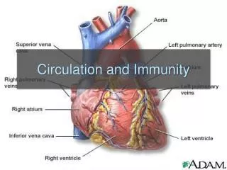
Heart Parts Continued Chambers • Atria – two cavities on top – right receives deoxygenated blood from body, left receives oxygenated blood from the lungs • Ventricles – the inferior and larger cavities – right pumps blood to the lungs, left pumps blood to the rest of body (*bigger ventricle and muscle is thicker as a result) • Septum – divides the heart into right and left
Heart Parts continued - Valves • Atrioventricular Valves (AV valves) (look like saloon doors) • Between the atria and ventricles • Allows for movement to blood from atria to ventricles • As the ventricles contract it forces the flap of the valve to close and open during ventricular relaxation • Left valve = bicuspid (2 flaps) or mitral valve • Right valve = tricuspid (3 flaps) • Semilunars(look like half moons) • Cover the pulmonary and aortic arteries • Open up into the artery • Open when the ventricles are contracting so that blood goes to the body or lungs • Back flow closes them so that blood doesn’t flow back into the heart
(aortic valve) (pulmonary valve) (tricuspid) (bicuspid or mitral valve)