Download
1 / 58
580 likes | 614 Views
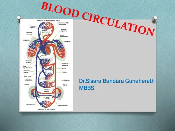
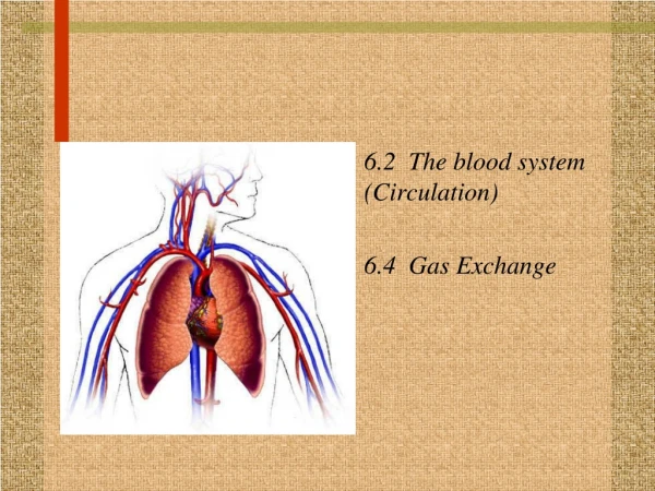
6.2 The blood system (Circulation) 6.4 Gas Exchange. Human Circulatory System. Essential idea: The blood system continuously transports substances to cells and simultaneously collects waste products. Human Circulatory System. Cardiovascular system •heart (atria/ventricles)

E N D
6.2 The blood system (Circulation) • 6.4 Gas Exchange
Human Circulatory System • Essential idea: The blood system continuously transports substances to cells and simultaneously collects waste products.
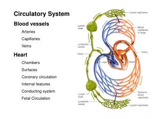
Human Circulatory System • Cardiovascular system • •heart (atria/ventricles) • •blood vessels (arteries, arterioles, capillary beds, venules, veins) • •blood (circulatory fluid)
The HEART Skill: Recognition of the chambers and valves of the heart and the blood vessels connected to it in dissected hearts or in diagrams of heart structure. (see next slide)
Draw and Label: show 4 chambers, associated blood vessels, valves and route of blood through heart • Parts to label: • Vena Cava (superior and inferior) • right atrium • Tricuspid valve (atrioventricular) • Right ventricle • Pulmonic valve (semilunar) • Pulmonary artery • Left atrium • Bicuspid/Mitral valve (atrioventricular) • Left Ventricle • Aortic valve (semilunar) • Aorta
Draw and Label: • Parts to label: • Vena Cava (superior and inferior) • right atrium • Tricuspid valve (atrioventricular) • Right ventricle • Pulmonic valve (semilunar) • Pulmonary artery • Left atrium • Bicuspid/Mitral valve (atrioventricular) • Left Ventricle • Aortic valve (semilunar) • Aorta
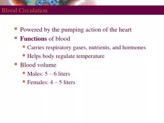
Heart Action • Atria collect blood from veins. • Contraction of atria pumps blood to ventricles through AV valves. • Contraction of ventricles pumps blood through semilunar valves into arteries. http://www.nhlbi.nih.gov/health/dci/Diseases/hhw/hhw_pumping.html
Important note: • There is a separate circulation for the lungs.
Application: Pressure changes in the left atrium, left ventricle and aorta during the cardiac cycle. • Discuss, then explain how and why these pressure changes occur in your notes…
Heartbeat Control • Myogenic muscle contraction: heart cells beat on their own. • Sinoatrial (SA) node (pacemaker)- a group of specialized muscle cells in the right atrium • Initiates heart beat by sending an electrical signal that propagates through the walls of the atria and then the walls of the ventricles. • Medulla of the brain– monitors and controls heartbeat. • The heart rate can be increased or decreased by impulses from two nerves from the medulla. • Epinephrine (adrenaline)– increases heart rate. http://www.nhlbi.nih.gov/health/dci/Diseases/hhw/hhw_electrical.html
Coronary arteries • Coronary arteries supply heart muscle with: • Oxygen • Nutrients
Application: Causes and consequences of occlusion of the coronary arteries. • Coronary occlusion = narrowing of coronary arteries • Causes/associations: • High blood concentrations of LDL. • trans fats consumption • Diabetes • Chronic high blood pressure. • Recent research indicates that certain infections can also play a role.
Application: Causes and consequences of occlusion of the coronary arteries. • Consequences: • Atherosclerosis (narrowing and hardening of arteries due to atheroma tissue) • Angina (pain due to oxygen deprivation etc.) • Heart attack What is an atheroma? An atheroma is a gathering of macrophage cells and debris in the artery walls. It is commonly described as an abnormal fatty accumulation in an artery.
Blood Vessels • 3 main types: Arteries, capillaries, and veins. • Relate structure to function: • Arteries: Have thick elastic fibres in their walls; thick smooth muscle • assist in maintaining blood pressure between pump cycles • Function: convey blood at high pressure from the ventricles to the tissues of the body
Blood vessels • Relate structure to function: • Capillaries • Permeable, thin-walled vessels • Function: Exchange materials between blood and tissues • Deliver oxygen and nutrients. • Pick up cell wastes • http://health.howstuffworks.com/adam-200084.htm
Blood vessels • Relate structure to function: • Veins • thin connective tissue; thin smooth muscle, valves (prevent backflow) • Function: collect blood at low pressure from the tissues of the body and return it to the atria of the heart • F:\Desktop\animations from bio powerpoints\Chapter 37 BDOL IC
Skill: Identification of blood vessels as arteries, capillaries or veins from the structure of their walls.
Blood • Function = Transport! • Carries: • nutrients • oxygen • carbon dioxide • hormones • antibodies • urea • heat
Blood Components • Plasma: liquid matrix of blood in which cells are suspended (90% water) • Erythrocytes (RBCs): transport O2 via hemoglobin (250 million per RBC?) • Leukocytes (WBCs): defense and immunity • Include phagocytes and lymphocytes • Platelets: clotting • Stem cells: pluripotent cells in the red marrow of bones • Blood clotting: fibrinogen (inactive)/ fibrin (active); hemophilia; thrombus (clot in blood vessels) • http://health.howstuffworks.com/adam-200028.htm
• Application: William Harvey’s discovery of the circulation of the blood with the heart acting as the pump. • Nature of science: Theories are regarded as uncertain—William Harvey overturned theories developed by the ancient Greek philosopher Galen on movement of blood in the body. • HW: Read about these on pages 290-291 of textbook or in Kognity. • Summarize in your notes A portrait of William Harvey
Utilization: • Understanding of the structure of the cardiovascular system has allowed the development of heart surgery. http://www.smm.org/heart/ Surgery video and other resources
Theory of knowledge: • Our current understanding is that emotions are the product of activity in the brain rather than the heart. Is knowledge based on science more valid than knowledge based on intuition?
Essential idea: The lungs are actively ventilated to ensure that gas exchange can occur passively. 6.4 Gas Exchange
Air is carried to the lungs in the trachea and bronchi and then to the alveoli in bronchioles Trachea (windpipe) Bronchi (tube to lungs) Bronchioles Alveoli (air sacs)– diffusion of gases due to differences in partial pressures. Diaphragm (breathing muscle) Respiratory system anatomy
Draw and Label Ventilation System • Include: • trachea • Lungs • Bronchi • Bronchioles • Alveoli (draw at high magnification as well) Students should be able to draw a diagram to show the structure of an alveolus and an adjacent capillary
Ventilation • maintains concentration gradients of oxygen and carbon dioxide between air in alveoli and blood flowing in adjacent capillaries. • Necessary for gas exchange.
Distinguish the following: • Ventilation: The act of breathing/ movement of air • Gas exchange: exchange of gases between blood and air. • Cell respiration: production of ATP in cells (why we breathe)
Mechanism of ventilation in Lungs Muscle contractions cause the pressure changes inside the thorax that force air in and out of the lungs to ventilate them. Different antagonistic muscles are required for inspiration and expiration because muscles only do work when they contract.
Mechanism of ventilation in Lungs Inhalation (Inspiration): breathing in • Volume of lungs increases • Pressure in lungs decreases • Negative pressure breathing: pulls air into lungs How? • diaphragm contracts • External intercostal muscles contract (lift ribcage) • Internal intercostal muscles relaxed • Abdominal muscles relaxed
Mechanism of ventilation in Lungs • Exhalation (Expiration): • Volume of lungs decreases • Pressure in lungs increases • Air is forced out How? • diaphragm relaxes • External intercostal muscles relaxed • Internal intercostal muscles contract (pull ribcage down and in) • Abdominal muscles contract (push up on diaphragm)
Gas exchange • Occurs across moist membranes. • CO2 diffuses into alveoli • O2 diffuses into blood • https://www.youtube.com/watch?v=d-f3RL0KiUg Overview ANIMATION
Alveoli • Adapted for gas exchange • Large surface area (many alveoli) • wall of alveoli = single layer of flattened cells (thin = good for gas exchange) • Moist film • Dense capillary network
Alveoli Cell Types • Type I pneumocytes • extremely thin alveolar cells • adapted to carry out gas exchange. • Type II pneumocytes • secrete a solution (ensures moisture) • Has surfactant that prevents the sides of the alveolus adhering to each other by reducing surface tension. (prevents alveolar collapse) Premature babies with insufficient surfactant may experience respiratory distress syndrome. Treated by giving oxygen and surfactant from animal lungs
Nature of science: • Obtain evidence for theories—epidemiological studies have contributed to our understanding of the causes of lung cancer. (1.8) • Epidemiology = the study of the incidence and causes of disease • Most studies are observational and statistical, rather than experimental in human populations. (Why?) • Looks for correlations/associations
Lung Cancer (Causes and Consequences) (note % shown is % of cases attributed to this cause) Causes: • Smoking (87%) • Asbestos, silica dust etc. • Passive smoking (secondhand smoke); about 3% of all cases. • Air pollution: exhaust and smoke. (about 5%) • Radon gas: leaks from certain rocks.
Lung Cancer (Causes and Consequences) (note % shown is % of cases attributed to this cause) Possible consequences: • High mortality rate • Shortness of breath. • Persistent Cough • Coughing up blood. • Pain (chest pain and pain in other areas if cancer spreads). • Accumulation of fluid in the chest. • Metastasis • Loss of appetite or weight loss. • Fatigue • Repeated problems with pneumonia or bronchitis.
Emphysema (Causes and Consequences) Causes: • long term exposure to cigarette smoke and other pollutants • Results: inflammation, narrowing of small airways and breakdown of lung tissue. • alveoli become less elastic • breakdown of alveolar wall — creating one larger air space instead of many small ones. Reduces lung surface area/gas exchange.
Emphysema (Causes and Consequences) Possible consequences: • Irreversible damage to lung • Shortness of breath. • Persistent cough. • Fatigue • Weight loss.
Skill: Monitoring of ventilation in humans at rest and after mild and vigorous exercise. (Practical 6) • Ventilation rate and tidal volume should be measured. • Ventilation rate: number of breaths taken during a specific amount of time. • Tidal volume: the amount of air which enters or leaves the lungs in a single breath at rest. (Average: 500 ml)
The heartbeat (option H) • Sinoatrial (SA) node (“pacemaker”): sets rate and timing of cardiac contraction by generating electrical signals • Atrioventricular (AV) node: relay point (0.1 second delay) spreading impulse to walls of ventricles • Electrocardiogram (ECG or EKG)
OPTION H Partial Pressure = the pressure exerted by each component in a mixture. • Gases will always flow from a region of higher partial pressure to one of lower pressure • The pressure of a gas in a mixture is the same as it would exert if it occupied the same volume alone at the same temperature (Dalton’s Law).
Respiratory pigments: gas transport • Oxygen transport- • Hemoglobin: vertebrates (Fe) • Carbon dioxide transport- • Blood plasma (7%) • Hemoglobin (23%) • MOST= Bicarbonate ions (HCO3- ) in plasma (70 -85%) • Carbonic anhydrase catalyses the following reaction: • H2O + CO2 H+ + HCO3- • Makes buffer system (Bicarbonate/ Carbonic Acid) preventing the blood from becoming too acidic
Option H: Explain the oxygen dissociation curves of adult and fetal hemoglobin and myoglobin. • As oxygen is bound, hemoglobin changes shape and has a higher affinity for binding more oxygen molecules. • Fetal Hemoglobin has a higher affinity for oxygen than the adult so that it can extract oxygen from the placenta. • What if the curve shifts right? Left? • As the curves shift to the right, hemoglobin releases oxygen more easily.
Option H: Explain the role of the Bohr Shift in the supply of oxygen to respiring tissues. • As CO2 increases, hemoglobin’s affinity for O2 decreases. • CO2 released in tissues causes the delivery of oxygen to increase to those tissues. • Why is this good?
Option H: Lung Cancer, Asthma, altitude • Lung cancer • Causes: smoking, because of the carcinogens in it. Other environmental carcinogens in lungs
Option H • Asthma • Causes: hereditary, attacks can be triggered by allergens like pollen and dust. It inflames and constricts the bronchial tubes which lead to wheezing and coughing
Option H • Altitude • Acclimatization • Increased RBC production • Increased ventilation rate • Altitude Sickness More info: http://www.ismmed.org/np_altitude_tutorial.htm
Breathing • Negative pressure breathing: pulls air into lungs (mammals) • Inhalation: diaphragm contraction; Exhalation: diaphragm relaxation • Tidal volume: amount of air inhaled and exhaled with each breath (500ml) • Vital capacity: maximum tidal volume during forced breathing (4L) • Regulation: CO2 concentration (as carbonic acid) in blood • Sensed by medulla oblongata, and chemosensors in aorta and carotid arteries (which relay the message to the brain)