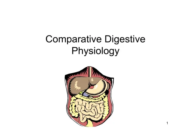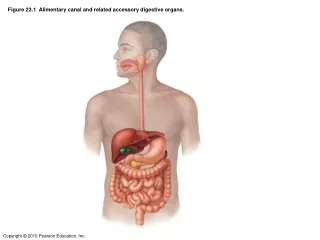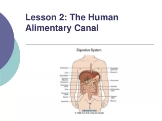
Digestive System: Alimentary Canal
170 likes | 423 Views
Digestive System: Alimentary Canal. Metallic 0 Mind. Submucosa : Dense irregular fibroelastic connvtive tissue. No glands Except in the esophagus and duodenum. Meissner’s submucosal plexus: houses postganglionic nerve cell bodies. Mucosa: Lined by epithelium.

Digestive System: Alimentary Canal
E N D
Presentation Transcript
Digestive System: Alimentary Canal Metallic 0 Mind
Submucosa: • Dense irregular fibroelasticconnvtive tissue. • No glands Except in the esophagus and duodenum. • Meissner’ssubmucosal plexus: houses postganglionic nerve cell bodies. • Mucosa: • Lined by epithelium. Deep is a loose connective tissue (Lamina Propria) that houses glands and lymph vessels. • Mucsularismucosaesurround lamina propria and composed of : • Inner circular layer. • Outer longitudinal layer
Connective tissue envelopes the muscularisexterna that may or may not surrounded by squamous epithelium If: • MuscularisExterna • Responsible for peristaltic activity. • Smooth muscle Except in the esophagus. • Interstitial cells of cajalare the pacemakers. • Arranged helically the organ is inraperitoneal it’s known as Serosa Composed of The organ is retroperitoneal it’s known as adventitia Between the muscle is auerbach’smyenteric plexus composed of postganglionic parasympatheric nerve cells.
Mucousa: Epithelium: Stratified squamousnonkeratinized epithelium. • Lamina propria: • Unremarkable. • Houses esophageal cardiac glands in two clusters: • Near the pharynx. • Near its juncture with stomach. MuscularisMucosae: Single layer of longitudinal smooth muscle.
Submusosa: Muscularisexterna • Inner circular and outer longitudinal layers. • They have both skeletal and smooth. • fibroelastic connective tissue. • Houses esophageal glands proper. In the upper third of esophagus it’s mostly skeletal In the middle third it’s both skeletal and smooth In the lowest third it’s only smooth The esophagus it covered by and adventitia until it pierces the diaphragm after that it’s covered by serosa.
Stomach • Rugae: • Longitudinal folds (transverse in the anturm) of mucosa and submucosa. • Disappear in distended stomach. • Gastric Pits: • Formed by epithelial lining. • Gastric glands empty in the bottom of each gastric pit.
Fundic Mucosa • Epithelium : • Simple columnar epithelium. • surface lining cells : manufacture mucin. • No goblet cells. • Lamina propria: • Connective tissue. • Occupied by Fundic (oxyntic) Glands. • Fundic Glands: • short pits. • Simple or branched. • Have 3 regions: • Isthmus. • Neck. • Base.
DNES cells (APUD or enteroendocrine cells) • In the base region. • Manufacture: endocrine, paracrine, neurocrine hormones. • Well-developed RER and golgi apparatus and numerous mitochondria. • Basal granules. • If: • Regenerative (stem) cells: • In the neck region. • Proliferate to replace all of the specialized cells • Parietal (oxyntic) Cells: • at the periphery of the gland. • Produce (HCL) and intrinsic factors. • Basally located nuclei. • Acidophilic • Apical membrane invaginates to form intracellular canaliculilined by microvilli. • Cytoplasm in the canaliculi has round and tubular vesicles tubulovesicular system. • Rich in mitochondria. • Chief (zymogenic) cells: • In the base region. • Manufacture pepsinogen, renin, gastric lipase • Exhibit rich RER, golgi apparatus, apical granules. • Basophilic. • Mucous neck cells: • In the neck region. • Secrete mucous to lubricate gastric content. • Golgi apparatus , RER are present. • Mitochondria in the basal region of the cell. • Apical cytoplasm has secretory granules. The cell reach the lumen of the gut called (the open type) The cell doesn’t reach the lumen called (the closed type)
Submucosa of the stomach: Dense, irregular collagenous connective tissue. MuscularisExterna: Three layers: Middle Circular muscle layer: Especially pronounced where it forms the pyloric sphincter. Outer longitudinal muscle layer Innermost Oblique muscle layer: Not well defined except in the cardiac region. • Serosa: • Thin loose subserous connective tissue. • Covered by wet smooth wet simple squamous epithelium.
Small Intestine 3 types of modification are present in the small intestine to increase the surface area: • PlicaeCirculares (valves of Kerckring) • Transverse folds of submucosa and mucosa. • Permanent. • Inncrease surface area by factor 2 to 3 • Villi: • Protrusions of the lamina propria. • Epithelially covered. • The core composed of : • Capillary loops • Lymphatic channel (lacteal) • Few smooth muscle fibers. • Loose connective tissue rich in lymphoid cells • Numbers are greater in the duodenum. • Increase the surface area 10 times Microvilli: Increase by factor of 20 Invaginations of the epithelium into the lamina propria between villi form glands (crypts of Lieberkühn)
Intestinal mucosa in regions where lymphoid nodules abut the epithelium. • M cells replace simple columnar epithelial lining of the small intestine. • Function: presents antigen
Lamina Propria Loose connective tissue. • Crypts of Lieberkuhn: • Tubular (or branched) gland. • Open into the intervillus space. • Paneth cells: • In the bottom of the crypts. • Acidophilic • Apical granules. • Manufacture lysozyme.
Muscularismucoasae: Inner circular layer. Outer longitudinal layer • Submucosa: • Irregular fibroelastic connective tissue. • Submucosa of the duodenum houses glands known as Brunner’s Glands. MuscularisExterna: Inner circular layer. Outer longitudinal layer. Second and third part of the duodenum have adventitia. Everything else has serosa.
Large Intestine • Colon Histology: • No villi. • Have crypts of lieberkühn. • Absent Paneth cells. Lamina propria, muscularis mucosa, submuosaresemble small intestine. • Muscularisexterna: • have unusual outer longitudinal muscle layer. • The muscle gathered in three narrow ribbons known as taenie coli Serosa has a fat-filled pouches called appendices epiploicae.
Appendix Vermiform appendix • Mucosa: • Simple columnar epithelium consisting of surface absorptive , goblet cells, M cells. • Lamina propria : loose connective tissue with lymph nodules and crypts of liebekühn • Same muscularismucosae as colon. • Same submucosa also it have lymphoid nodules and fatty infiltration. • Same muscularisexterna. • Covered by serosa
Please Remember Meissner’s plexus and Auerbach plexus present in all tissues of alimentary canal. Goblet cells starts at the duodenum and increases gradually