Download
1 / 78
800 likes | 1.16k Views
Explore the intricate workings and structures of the gastrointestinal tract, including peristalsis, segmentation, and neural reflex pathways. Learn about the peritoneum, mucosa, and oral cavity anatomy.

E N D
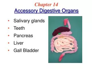
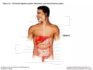
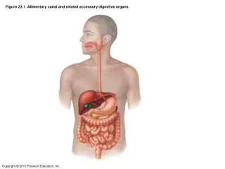
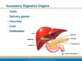
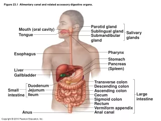
Figure 23.1 Alimentary canal and related accessory digestive organs. Parotid gland Mouth (oral cavity) Sublingual gland Salivary glands Tongue Submandibular gland Pharynx Esophagus Stomach Pancreas (Spleen) Liver Gallbladder Transverse colon Duodenum Descending colon Jejunum Small intestine Ascending colon Large intestine Ileum Cecum Sigmoid colon Rectum Vermiform appendix Anal canal Anus
Figure 23.2 Gastrointestinal tract activities. Ingestion Food Mechanical Pharynx digestion Esophagus • Chewing (mouth) Propulsion • Churning (stomach) • Segmentation (small intestine) • Swallowing (oropharynx) Chemical • Peristalsis (esophagus, stomach, small intestine, large intestine) digestion Stomach Absorption Lymph vessel Small intestine Blood vessel Large intestine Mainly H2O Feces Anus Defecation
Figure 23.3 Peristalsis and segmentation. From mouth (b) Segmentation: Nonadjacent segments of alimentary tract organs alternately contract and relax, moving the food forward then backward. Food mixing and slow food propulsion occurs. (a) Peristalsis: Adjacent segments of alimentary tract organs alternately contract and relax, which moves food along the tract distally.
Figure 23.3a Peristalsis and segmentation. From mouth (a) Peristalsis: Adjacent segments of alimentary tract organs alternately contract and relax, which moves food along the tract distally.
Figure 23.3b Peristalsis and segmentation. (b) Segmentation: Nonadjacent segments of alimentary tract organs alternately contract and relax, moving the food forward then backward. Food mixing and slow food propulsion occurs.
Figure 23.4 Neural reflex pathways initiated by stimuli inside or outside the gastrointestinal tract. External stimuli (sight, smell, taste, thought of food) Central nervous system and extrinsic autonomic nerves Long reflexes Afferent impulses Efferent impulses Effectors: Smooth muscle or glands Chemoreceptors, osmoreceptors, or mechanoreceptors Local (intrinsic) nerve plexus (“gut brain”) Internal (GI tract) stimuli Short reflexes Response: Change in contractile or secretory activity Gastrointestinal wall (site of short reflexes) Lumen of the alimentary canal
Figure 23.5 The peritoneum and the peritoneal cavity. Abdominopelvic cavity Vertebra Dorsal mesentery Parietal peritoneum Mesentery resorbed and lost Ventral mesentery Visceral peritoneum Peritoneal cavity Alimentary canal organ Liver (a) Schematic cross sections of abdominal cavity illustrate the peritoneums and mesenteries. Alimentary canal organ Alimentary canal organ in a retroperitoneal position (b) Some organs lose their mesentery and become retroperitoneal during development.
Figure 23.5a The peritoneum and the peritoneal cavity. Abdominopelvic cavity Vertebra Dorsal mesentery Parietal peritoneum Ventral mesentery Visceral peritoneum Peritoneal cavity Alimentary canal organ Liver (a) Schematic cross sections of abdominal cavity illustrate the peritoneums and mesenteries.
Figure 23.5b The peritoneum and the peritoneal cavity. Mesentery resorbed and lost Abdominopelvic cavity Alimentary canal organ Alimentary canal organ in a retroperitoneal position (b) Some organs lose their mesentery and become retroperitoneal during development.
Figure 23.6 Basic structure of the alimentary canal. Intrinsic nerve plexuses • Myenteric nerve plexus • Submucosal nerve plexus Glands in submucosa Mucosa • Epithelium • Lamina propria • Muscularis mucosae Submucosa Muscularis externa • Longitudinal muscle • Circular muscle Serosa Nerve • Epithelium Artery • Connective tissue Vein Lumen Gland in mucosa Lymphatic vessel Mucosa-associated lymphoid tissue Duct of gland outside alimentary canal Mesentery
Figure 23.7 Anatomy of the oral cavity (mouth). Soft palate Palatoglossal arch Uvula Hard palate Upper lip Gingivae (gums) Superior labial frenulum Oral cavity Palatine raphe Palatoglossal arch Palatine tonsil Hard palate Palatopharyngeal arch Tongue Soft palate Oropharynx Uvula Lingual tonsil Posterior wall of oropharynx Palatine tonsil Epiglottis Tongue Hyoid bone Sublingual fold with openings of sublingual ducts Lingual frenulum Laryngopharynx Opening of submandibular duct Gingivae (gums) Esophagus Vestibule Inferior labial frenulum Lower lip Trachea (b) Anterior view (a) Sagittal section of the oral cavity and pharynx
Figure 23.7a Anatomy of the oral cavity (mouth). Soft palate Palatoglossal arch Uvula Hard palate Oral cavity Palatine tonsil Tongue Oropharynx Lingual tonsil Epiglottis Hyoid bone Laryngopharynx Esophagus Trachea (a) Sagittal section of the oral cavity and pharynx
Figure 23.7b Anatomy of the oral cavity (mouth). Upper lip Gingivae (gums) Superior labial frenulum Palatine raphe Palatoglossal arch Hard palate Palatopharyngeal arch Soft palate Uvula Posterior wall of oropharynx Palatine tonsil Tongue Sublingual fold with openings of sublingual ducts Lingual frenulum Opening of submandibular duct Gingivae (gums) Vestibule Inferior labial frenulum Lower lip (b) Anterior view
Figure 23.8 Dorsal surface of the tongue, and the tonsils. Epiglottis Palatopharyngeal arch Palatine tonsil Lingual tonsil Palatoglossal arch Terminal sulcus Foliate papillae Circumvallate papilla Midline groove of tongue Dorsum of tongue Fungiform papilla Filiform papilla
Figure 23.9 The salivary glands. Ducts of sublingual gland Tongue Teeth Parotid gland Parotid duct Masseter muscle Lingual frenulum Body of mandible (cut) Sublingual gland Posterior belly of digastric muscle Mylohyoid muscle (cut) Submandibular duct Mucous cells Serous cells forming demilunes Anterior belly of digastric muscle Submandibular gland (a) (b)
Figure 23.9a The salivary glands. Ducts of sublingual gland Tongue Teeth Parotid gland Parotid duct Masseter muscle Lingual frenulum Body of mandible (cut) Sublingual gland Posterior belly of digastric muscle Mylohyoid muscle (cut) Submandibular duct Anterior belly of digastric muscle Submandibular gland (a)
Figure 23.9b The salivary glands. Mucous cells Serous cells forming demilunes (b)
Figure 23.10a Human dentition. Incisors Incisors Central (7 yr) Central (6–8 mo) Lateral (8 yr) Lateral (8–10 mo) Canine (eyetooth) (11 yr) Canine (eyetooth) (16–20 mo) Premolars (bicuspids) Molars First molar (10–15 mo) First premolar (11 yr) Deciduous (milk) teeth Second molar (about 2 yr) Second premolar (12–13 yr) Molars First molar (6–7 yr) Second molar (12–13 yr) Third molar (wisdom tooth) (17–25 yr) Permanent teeth (a)
Figure 23.10b Human dentition. (b) Deciduous teeth Permanent teeth
Figure 23.11 Longitudinal section of a canine tooth within its bony alveolus. Enamel Dentin Crown Dentinal tubules Pulp cavity (contains blood vessels and nerves) Neck Gingiva (gum) Cementum Root canal Root Periodontal ligament Apical foramen Bone
Figure 23.12 Microscopic structure of the esophagus. Mucosa (contains a stratified squamous epithelium) Submucosa (areolar connective tissue) Lumen Muscularis externa • Circular layer • Longitudinal layer Adventitia (fibrous connective tissue) (a) (b)
Figure 23.12a Microscopic structure of the esophagus. Mucosa (contains a stratified squamous epithelium) Submucosa (areolar connective tissue) Lumen Muscularis externa • Longitudinal layer • Circular layer Adventitia (fibrous connective tissue) (a)
Figure 23.12b Microscopic structure of the esophagus. Mucosa (contains a stratified squamous epithelium) (b)
Figure 23.13 Deglutition (swallowing) (1 of 5). Bolus of food Tongue Pharynx Epiglottis Glottis Trachea 1 Upper esophageal sphincter is contracted. During the buccal phase, the tongue presses against the hard palate, forcing the food bolus into the oropharynx where the involuntary phase begins.
Figure 23.13 Deglutition (swallowing) (2 of 5). Uvula Bolus Epiglottis Esophagus 2 The uvula and larynx rise to prevent food from entering respiratory passageways. The tongue blocks off the mouth. The upper esophageal sphincter relaxes, allowing food to enter the esophagus.
Figure 23.13 Deglutition (swallowing) (3 of 5). Bolus 3 The constrictor muscles of the pharynx contract, forcing food into the esophagus inferiorly. The upper esophageal sphincter contracts (closes) after entry.
Figure 23.13 Deglutition (swallowing) (4 of 5). Relaxed muscles 4 Food is moved through the esophagus to the stomach by peristalsis. Circular muscles contract Bolus of food Longitudinal muscles contract Gastroesophageal sphincter closed Stomach
Figure 23.13 Deglutition (swallowing) (5 of 5). 5 The gastroesophageal sphincter opens, and food enters the stomach. Relaxed muscles Gastroesophageal sphincter opens
Figure 23.14a Anatomy of the stomach. Cardia Fundus Esophagus Muscularis externa Serosa • Longitudinal layer • Circular layer Body • Oblique layer Lumen Lesser curvature Rugae of mucosa Greater curvature Pyloric canal Pyloric antrum Duodenum Pyloric sphincter (valve) at pylorus (a)
Figure 23.14b Anatomy of the stomach. Fundus Liver (cut) Body Spleen Lesser curvature Greater curvature (b)
Figure 23.15a Microscopic anatomy of the stomach. Surface epithelium Mucosa Lamina propria Muscularis mucosae Submucosa (contains submucosal plexus) Oblique layer Muscularis externa (contains myenteric plexus) Circular layer Longitudinal layer Serosa Stomach wall (a) Layers of the stomach wall (l.s.)
Figure 23.15b Microscopic anatomy of the stomach. Gastric pits Surface epithelium (mucous cells) Gastric pit Mucous neck cells Parietal cell Chief cell Gastric gland Enteroendocrine cell (b) Enlarged view of gastric pits and gastric glands
Figure 23.15c Microscopic anatomy of the stomach. Pepsinogen Pepsin HCl Mitochondria Parietal cell Chief cell Enteroendocrine cell (c) Location of the HCl-producing parietal cells and pepsin-secreting chief cells in a gastric gland
Figure 23.16 Photographs of a gastric ulcer lesion and of the bacteria that most commonly cause it. Bacteria Mucosa layer of stomach (b) H. pylori bacteria (a) A gastric ulcer lesion
Figure 23.16a Photographs of a gastric ulcer lesion and of the bacteria that most commonly cause it.
Figure 23.16b Photographs of a gastric ulcer lesion and of the bacteria that most commonly cause it. Bacteria Mucosa layer of stomach (b) H. pylori bacteria
Figure 23.17 Neural and hormonal mechanisms that regulate release of gastric juice. Stimulatory events Inhibitory events 1 1 Sight and thought of food Loss of appetite, depression Cerebral cortex Lack of stimulatory impulses to parasym- pathetic center Cerebral cortex Cephalic phase Conditioned reflex 2 Stimulation of taste and smell receptors Hypothalamus and medulla oblongata Vagus nerve 1 1 Stomach distension activates stretch receptors Excessive acidity (pH <2) in stomach Vagovagal reflexes Medulla Vagus nerve Gastrin secretion declines G cells Gastric phase 2 Emotional upset Local reflexes Overrides parasym- pathetic controls Sympathetic nervous system activation 2 Food chemicals (especially peptides and caffeine) and rising pH activate chemoreceptors G cells Gastrin release to blood Stomach secretory activity 1 Distension of duodenum; presence of fatty, acidic, hypertonic chyme, and/or irritants in the duodenum Entero- gastric reflex Local reflexes 1 Presence of low pH, partially digested foods, fats, or hypertonic solution in duodenum when stomach begins to empty Intestinal (enteric) gastrin release to blood Vagal nuclei in medulla Brief effect Intestinal phase Pyloric sphincter 2 Distension; presence of fatty, acidic, partially digested food in the duodenum Release of intestinal hormones (secretin, cholecystokinin, vasoactive intestinal peptide) Stimulate Inhibit
Figure 23.18 Mechanism of HCl secretion by parietal cells. Blood capillary Chief cell Stomach lumen CO2 CO2 + H2O H+-K+ ATPase Carbonic anhydrase H2CO3 H+ H+ K+ K+ HCO3– HCI Parietal cell Alkaline tide HCO3– Cl– Cl– Cl– l HCO3–- Cl– antiporter Inter- stitial fluid
Figure 23.19 Peristaltic waves in the stomach. Pyloric valve closed Pyloric valve closed Pyloric valve slightly opened Propulsion: Peristaltic waves move from the fundus toward the pylorus. Grinding: The most vigorous peristalsis and mixing action occur close to the pylorus. Retropulsion: The pyloric end of the stomach acts as a pump that delivers small amounts of chyme into the duodenum, simultaneously forcing most of its contained material backward into the stomach. 1 2 3
Figure 23.20 Neural and hormonal factors inhibiting gastric emptying. Presence of fatty, hypertonic, acidic chyme in duodenum Duodenal entero- endocrine cells Chemoreceptors and stretch receptors Secrete Target Via short reflexes Via long reflexes Enterogastrones (secretin, cholecystokinin, vasoactive intestinal peptide) Enteric neurons CNS centers sympathetic activity; parasympathetic activity Duodenal stimuli decline Contractile force and rate of stomach emptying decline Initial stimulus Physiological response Stimulate Inhibit Result
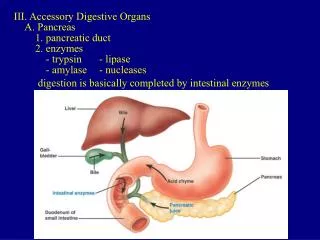
Figure 23.21 The duodenum of the small intestine, and related organs. Right and left hepatic ducts of liver Cystic duct Common hepatic duct Bile duct and sphincter Accessory pancreatic duct Mucosa with folds Tail of pancreas Pancreas Gallbladder Jejunum Major duodenal papilla Main pancreatic duct and sphincter Hepatopancreatic ampulla and sphincter Duodenum Head of pancreas
Figure 23.22a Structural modifications of the small intestine that increase its surface area for digestion and absorption. Vein carrying blood to hepatic portal vessel Muscle layers Lumen Circular folds Villi (a)
Figure 23.22b Structural modifications of the small intestine that increase its surface area for digestion and absorption. Microvilli (brush border) Absorptive cells Lacteal Goblet cell Vilus Blood capillaries Mucosa associated lymphoid tissue Enteroendocrine cells Intestinal crypt Venule Muscularis mucosae Lymphatic vessel Duodenal gland Submucosa (b)
Figure 23.22c Structural modifications of the small intestine that increase its surface area for digestion and absorption. Absorptive cells Goblet cells Villi (c) Intestinal crypt
Figure 23.23 Villi and microvilli of the small intestine. Villi Microvilli Desquamating cells Absorptive cell (a) (b)
Figure 23.23a Villi and microvilli of the small intestine. Villi Desquamating cells (a)
Figure 23.23b Villi and microvilli of the small intestine. Microvilli Absorptive cell (b)
Figure 23.24a Gross anatomy of the human liver. Sternum Bare area Nipple Falciform ligament Liver Left lobe of liver Right lobe of liver Round ligament (ligamentum teres) Gallbladder (a)
Figure 23.24b Gross anatomy of the human liver. Sternum Nipple Liver Bare area Lesser omentum (in fissure) Caudate lobe of liver Left lobe of liver Sulcus for inferior vena cava Porta hepatis containing hepatic artery (left) and hepatic portal vein (right) Hepatic vein (cut) Bile duct (cut) Right lobe of liver Quadrate lobe of liver Gallbladder Ligamentum teres (b)
Figure 23.25a-b Microscopic anatomy of the liver. (a) (b) Lobule Central vein Connective tissue septum