Download
1 / 71
710 likes | 846 Views
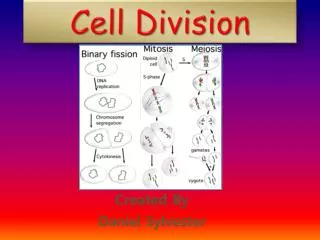
Cell Division. Mitosis and Meiosis. Binary Fission. Human Life Cycle. Cellular Organization of the Genetic Material. All the DNA in a cell constitutes the cell’s genome

E N D
Cell Division Mitosis and Meiosis
Cellular Organization of the Genetic Material • All the DNA in a cell constitutes the cell’s genome • A genome can consist of a single DNA molecule (common in prokaryotic cells) or a number of DNA molecules (common in eukaryotic cells) • DNA molecules in a cell are packaged into chromosomes
Every eukaryotic species has a characteristic number of chromosomes in each cell nucleus • Somatic cells (nonreproductive cells) have two sets of chromosomes • Gametes (reproductive cells: sperm and eggs) have half as many chromosomes as somatic cells • Eukaryotic chromosomes consist of chromatin, a complex of DNA and protein that condenses during cell division
Distribution of Chromosomes During Eukaryotic Cell Division • In preparation for cell division, DNA is replicated and the chromosomes condense • Each duplicated chromosome has two sister chromatids, which separate during cell division • The centromereis the narrow “waist” of the duplicated chromosome, where the two chromatids are most closely attached
0.5 µm Chromosomes DNA molecules Chromo- some arm Chromosome duplication (including DNA synthesis) Centromere Sister chromatids Separation of sister chromatids Centromere Sister chromatids
Eukaryotic cell division consists of: • Mitosis, the division of the nucleus • Cytokinesis, the division of the cytoplasm • Gametes are produced by a variation of cell division called meiosis • Meiosis yields nonidentical daughter cells that have only one set of chromosomes, half as many as the parent cell
Phases of the Cell Cycle • The cell cycle consists of • Mitotic (M) phase (mitosis and cytokinesis) • Interphase (cell growth and copying of chromosomes in preparation for cell division) • Interphase (about 90% of the cell cycle) can be divided into subphases: • G1 phase (“first gap”) • S phase (“synthesis”) • G2 phase (“second gap”) • The cell grows during all three phases, but chromosomes are duplicated only during the S phase
Fig. 12-5 INTERPHASE S (DNA synthesis) G1 Cytokinesis G2 Mitosis MITOTIC (M) PHASE
Phases of Mitosis BioFlix: Mitosis • Mitosis is conventionally divided into four or five phases: • Prophase • Prometaphase • Metaphase • Anaphase • Telophase • Cytokinesis is well underway by late telophase
Phases of Mitosis Metaphase Anaphase Telophase and Cytokinesis G2 of Interphase Prophase Prometaphase Centrosomes (with centriole pairs) Early mitotic spindle Centromere Chromatin (duplicated) Fragments of nuclear envelope Nonkinetochore microtubules Aster Cleavage furrow Metaphase plate Nucleolus forming Daughter chromosomes Nuclear envelope forming Centrosome at one spindle pole Spindle Nuclear envelope Kinetochore Chromosome, consisting of two sister chromatids Kinetochore microtubule Plasma membrane Nucleolus
Fig. 12-6b G2 of Interphase Prophase Prometaphase Chromatin (duplicated) Centrosomes (with centriole pairs) Early mitotic spindle Fragments of nuclear envelope Centromere Aster Nonkinetochore microtubules Kinetochore Nuclear envelope Plasma membrane Chromosome, consisting of two sister chromatids Kinetochore microtubule Nucleolus
Fig. 12-6d Telophase and Cytokinesis Metaphase Anaphase Nucleolus forming Metaphase plate Cleavage furrow Daughter chromosomes Nuclear envelope forming Centrosome at one spindle pole Spindle
Cytokinesis • In animal cells, cytokinesis occurs by a process known as cleavage, forming a cleavage furrow • In plant cells, a cell plate forms during cytokinesis
Fig. 12-UN2 Mitosis in plant cells
INTERPHASE Mitosis Review G1 S Cytokinesis Mitosis G2 MITOTIC (M) PHASE Prophase Telophase and Cytokinesis Prometaphase Anaphase Metaphase
Most animal cells exhibit density-dependent inhibition, in which crowded cells stop dividing • Most animal cells also exhibit anchorage dependence, in which they must be attached to a substratum in order to divide • Cancer cells exhibit neither density-dependent inhibition nor anchorage dependence
Fig. 12-19 Anchorage dependence Density-dependent inhibition Density-dependent inhibition 25 µm 25 µm (a) Normal mammalian cells (b) Cancer cells
Loss of Cell Cycle Controls in Cancer Cells • Cancer cells do not respond normally to the body’s control mechanisms • Cancer cells may not need growth factors to grow and divide: • They may make their own growth factor • They may convey a growth factor’s signal without the presence of the growth factor • They may have an abnormal cell cycle control system
A normal cell is converted to a cancerous cell by a process called transformation • Cancer cells form tumors, masses of abnormal cells within otherwise normal tissue • If abnormal cells remain at the original site, the lump is called a benign tumor • Malignant tumors invade surrounding tissues and can metastasize, exporting cancer cells to other parts of the body, where they may form secondary tumors
Fig. 12-20 Lymph vessel Tumor Blood vessel Cancer cell Glandular tissue Metastatic tumor Cancer cells invade neigh- boring tissue. A tumor grows from a single cancer cell. Cancer cells spread to other parts of the body. Cancer cells may survive and establish a new tumor in another part of the body. 4 2 1 3
Cell Division And Reproduction And Meiosis
Inheritance of Genes • Genes are the units of heredity, and are made up of segments of DNA • Genes are passed to the next generation through reproductive cells called gametes (sperm and eggs) • Each gene has a specific location called a locus on a certain chromosome • One set of chromosomes is inherited from each parent
Comparison of Asexual and Sexual Reproduction • In asexual reproduction, one parent produces genetically identical offspring by mitosis • A clone is a group of genetically identical individuals from the same parent • In sexual reproduction, two parents give rise to offspring that have unique combinations of genes inherited from the two parents
Sets of Chromosomes in Human Cells • Human somatic cells (any cell other than a gamete) have 23 pairs of chromosomes • A karyotype is an ordered display of the pairs of chromosomes from a cell • The two chromosomes in each pair are called homologous chromosomes, or homologs • Chromosomes in a homologous pair carry genes controlling the same inherited characters
Fig. 13-3 APPLICATION TECHNIQUE 5 µm Pair of homologous replicated chromosomes Centromere Sister chromatids Metaphase chromosome
Autosomes and Sex chromosomes • The sex chromosomes are called X and Y • Human females have a homologous pair of X chromosomes (XX) • Human males have one X and one Y chromosome • The 22 pairs of chromosomes that do not determine sex are calledautosomes
Each pair of homologous chromosomes includes one chromosome from each parent • The 46 chromosomes in a human somatic cell are two sets of 23: one from the mother and one from the father • A diploid cell (2n) has two sets of chromosomes • For humans, the diploid number is 46 (2n = 46)
Haploid cells • A gamete (sperm or egg) contains a single set of chromosomes, and is haploid (n) • For humans, the haploid number is 23 (n = 23) • Each set of 23 consists of 22 autosomes and a single sex chromosome
Review • In a cell in which DNA synthesis has occurred, each chromosome is replicated • Each replicated chromosome consists of two identical sister chromatids
Fig. 13-4 Diploid Cell Key Maternal set of chromosomes (n = 3) 2n = 6 Paternal set of chromosomes (n = 3) Two sister chromatids of one replicated chromosome Centromere Two nonsister chromatids in a homologous pair Pair of homologous chromosomes (one from each set)
Meiosis reduces the number of chromosome sets from diploid to haploid • Like mitosis, meiosis is preceded by the replication of chromosomes • Meiosis takes place in two sets of cell divisions, called meiosis I and meiosis II • The two cell divisions result in four daughter cells, rather than the two daughter cells in mitosis • Each daughter cell has only half as many chromosomes as the parent cell
The Stages of Meiosis (overview) • In the first cell division (meiosis I), homologous chromosomes separate • Meiosis I results in two haploid daughter cells with replicated chromosomes; it is called the reductional division • In the second cell division (meiosis II), sister chromatids separate • Meiosis II results in four haploid daughter cells with unreplicated chromosomes
Fig. 13-7-1 Interphase Homologous pair of chromosomes in diploid parent cell Meiosis overview Chromosomes replicate Homologous pair of replicated chromosomes Sister chromatids Diploid cell with replicated chromosomes
Fig. 13-7-2 Interphase Homologous pair of chromosomes in diploid parent cell Meiosis overview Chromosomes replicate Homologous pair of replicated chromosomes Sister chromatids Diploid cell with replicated chromosomes Meiosis I Homologous chromosomes separate 1 Haploid cells with replicated chromosomes
Fig. 13-7-3 Interphase Homologous pair of chromosomes in diploid parent cell Meiosis overview Chromosomes replicate Homologous pair of replicated chromosomes Sister chromatids Diploid cell with replicated chromosomes Meiosis I Homologous chromosomes separate 1 Haploid cells with replicated chromosomes Meiosis II Sister chromatids separate 2 Haploid cells with unreplicated chromosomes
Meiosis I is preceded by interphase, in which chromosomes are replicated to form sister chromatids • The sister chromatids are genetically identical and joined at the centromere BioFlix: Meiosis
Meiosis I Phases • Division in meiosis I occurs in four phases: – Prophase I – Metaphase I – Anaphase I – Telophase I and cytokinesis
Fig. 13-8b Prophase I Metaphase I Centrosome (with centriole pair) Centromere (with kinetochore) Sister chromatids Chiasmata Spindle Metaphase plate Homologous chromosomes Fragments of nuclear envelope Microtubule attached to kinetochore
Prophase I • Chromosomes begin to condense • In synapsis, homologous chromosomes loosely pair up, aligned gene by gene • Each pair of chromosomes forms a tetrad, a group of four chromatids • In crossing over, nonsisterchromatids exchange DNA segments • Each tetrad usually has one or more chiasmata, X-shaped regions where crossing over occurred
Metaphase I • In metaphase I, tetrads line up at the metaphase plate, with one chromosome facing each pole • Microtubules from one pole are attached to the kinetochore of one chromosome of each tetrad • Microtubules from the other pole are attached to the kinetochore of the other chromosome
Anaphase I • In anaphase I, pairs of homologous chromosomes separate • One chromosome moves toward each pole, guided by the spindle apparatus • Sister chromatids remain attached at the centromere and move as one unit toward the pole
Telophase I and Cytokinesis • In the beginning of telophase I, each half of the cell has a haploid set of chromosomes; each chromosome still consists of two sister chromatids • Cytokinesis usually occurs simultaneously, forming two haploid daughter cells • In animal cells, a cleavage furrow forms; in plant cells, a cell plate forms • No chromosome replication occurs between the end of meiosis I and the beginning of meiosis II because the chromosomes are already replicated
Meiosis II Phases • Division in meiosis II also occurs in four phases: – Prophase II – Metaphase II – Anaphase II –Telophase II and cytokinesis • Meiosis II is very similar to mitosis
Prophase II • In prophase II, a spindle apparatus forms • In late prophase II, chromosomes (each still composed of two chromatids) move toward the metaphase plate
Metaphase II • In metaphase II, the sister chromatids are arranged at the metaphase plate • Because of crossing over in meiosis I, the two sister chromatids of each chromosome are no longer genetically identical • The sister chromatids attach to microtubules extending from opposite poles
Anaphase II • In anaphase II, the sister chromatidsseparate • The sister chromatids of each chromosome now move as two newly individual chromosomes toward opposite poles