Download
1 / 44
460 likes | 1.02k Views
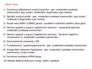
Otázky k testu:. Duchennova/Beckerova svalová dystrofie - gen, molekulární podstata onemocnění, typy mutací, molekulární diagnostika a její metody. Spinální svalová atrofie - gen, molekulární podstata onemocnění, typy mutací, molekulární diagnostika a její metody.

E N D
Otázky k testu: • Duchennova/Beckerova svalová dystrofie - gen, molekulární podstata onemocnění, typy mutací, molekulární diagnostika a její metody. • Spinální svalová atrofie - gen, molekulární podstata onemocnění, typy mutací, molekulární diagnostika a její metody. • Rozdíl mezi SMN1 a SMN2 genem, vysvětlení rozdílného sestřihu obou genů. • Nemoci spojené s expanzí repetitivních sekvencí - myotonická dystrofie, molekulární podstata onemocnění. • Nemoci spojené s expanzí repetitivních sekvencí - Syndrom fragilního chromozomu X, molekulární podstata onemocnění. • Novorozenecký screening v ČR. • Fenylketonurie, hyperfenylalaninemie - gen, molekulární podstata onemocnění. • Kongenitální adrenální hyperplazie - gen, molekulární podstata onemocnění, mechanizmy vzniku mutací. • Nonsense mediated mRNA decay • Metody detekce bodových mutací, delecí, duplikací
Duchennova svalová dystrofie (DMD) Incidence: 1/3500 chlapců; Mutace v genu pro dystrofin (DMD, Xp21); dystrofin - protein membránového skeletu svalových buněk, zajišťuje stabilitu svalové membrány
Molekulárně genetická diagnostika: analýza DNA Analýza 79 exonů metodou MLPA - Multiple Ligation dependent Probe Amplification Izolace DNA (leukocyty) Delece/duplikace není detekována Analýza jednotlivých exonů na přítomnost bodových mutací (PCR-sekvenování) Delece/duplikace je detekována (cca 60% pacientů) Určení přenašečství delece/duplikace v rodině
Na úrovni DNA delece/duplikace není detekována → imunohistochemická analýza dystrofinu ve svalu Normální vzorek BMD DMD
Molekulárně genetická diagnostika: analýza mRNA Izolace RNA (svalové buňky) Reverzní transkripce, amplifikace (10 fragmentů) Delece/duplikace není detekována Elektroforéza Protein truncation test - specifická detekce nonsense a frame-shift mutací Delece/duplikace je detekována M K P1 P2 P3 P4
Detekce rozsáhlých delecí/duplikací – Multiplex Ligation-dependent Probe Amplification • Denatured genomic DNA is hybridised with a mixture of probes. Each MLPA probe consists of two oligonucleotides. • Oligonucleotides hybridise to adjacent target sequences and are ligated by a thermostable ligase. • All probe ligation products are amplified simultaneously by PCR using a single primer pair. The amplification product of each probe has a unique length. • Amplification products are separated by capillary electrophoresis. Relative amounts of probe amplification products reflect the relative copy number of target sequences.
Reverzní transkripce cDNA 1. PCR PCR produkt 2. PCR T7 ATG PCR produkt In vitro transkripce/translace TAG RNA Protein Protein truncation test mRNA K P
Duchennova svalová dystrofie - nonsense mutace, delece a duplikace měnící čtecí rámec translace Beckerova svalová dystrofie - delece a duplikace zachovávající čtecí rámec translace Imunodetekce dystrofinu pomocí protilátky DYS2 ve svalové tkáni Normální vzorek Pacient s BMD Western blot Pacient s DMD
Spinální svalová atrofie (SMA) • Incidence: 1/6000 • Incidence přenašečů onemocnění: 1/50 • Charakterizována degradací alfa-motorických neuronů míchy a atrofií svalů. • Onemocnění s autozomálně recesivním typem dědičnosti. • 95% SMA je způsobeno homozygotní delecí genu SMN1 (Survival of Motor Neuron 1), který je lokalizován na chromozomu 5q12-5q13.38. Tato oblast obsahuje invertovanou duplikaci - SMN1 a SMN2 gen. • V důsledku rekombinace (crossing-over a genová konverze) vznikají delece, duplikace, změny počtu kopií SMN genů, ………..
Human Molecular Genetics, 2010, Vol. 19, Review Issue 1, R111–R118
SMN1: TTC (Phe) SMN2: TTT(Phe) • v 95% vzniká mRNA obsahující všechny exony • v 85% vzniká mRNA s delecí exonu 7 • v 10% vzniká mRNA obsahují všechny exony
SMN protein has a ubiquitous and essential function involving production ofsmall nuclear ribonucleoprotein complexes - active in pre-mRNA splicing.
del del K1 SMN1 SMN2 K2 Stanovení delece genu SMN1 • PCR a restrikční analýza (stanovení homozygotní delece SMN1 - sledování přítomnosti 7. exonu) • SMN1 gen: 188 pb, SMN2 gen: 149 pb + 39 pb • SALSA MLPA kit P060 (stanovení homozygotní delece SMN1 + stanovení počtu kopií SMN1) • Real-time PCR • (stanovení homozygotní delece SMN1 + stanovení počtu kopií SMN1)
RNA Biology 7:4, 430-440; July/August 2010 Splicing architecture of exon 7 of the human SMN1 and SMN2 genes. The diagram represents exon 7 (yellow box) and its flanking intronic regions (lines). Elements inhibiting exon 7 inclusion are shown in red, whereas the positive elements are represented in dark blue. The suboptimal branch point (BP) and polypyrimidine tract (PP tract) are indicated in light blue. SF2/ASF and Tra2/β1 bind to the exonic splicing enhancers SE1 and SE2, respectively. The recognition of SE1 by SF2/ASF is prevented in SMN2, due to the C → U transition. This sequence alteration also creates a hnRNP A1-dependent splicing silencer. Exon 7 is extremely short (only 54 bp).
a | The essential splicing signals that define the exon boundaries - GU and AG dinucleotides, the branch-point adenosine, polypyrimidine tract of variable length. Components of the basal splicing machinery bind to the consensus sequences and promote assembly of the splicing complex. The U1 snRNP binds to the 5'-splice site, and the U2 snRNP binds the branch site through RNA–RNA interactions. Additional enhancer and silencer elements in the exons and introns (ESE, ESS, ISE, ISS) allow the correct splice sites to be distinguished from the many cryptic splice sites that have identical signal sequences. Trans-acting splicing factors can interact with enhancers and silencers and can accordingly be subdivided into two main groups: members of the SR family of proteins and of the hnRNPs. In general, SR protein binding at ESE facilitates exon recognition whereas hnRNPs are inhibitory. Protein–protein interactions in the spliceosome that modulate the recognition of the splice sites are the probable cause of splicing inhibition or activation. b | Genomic variants (GVs) can affect different splicing regulatory elements, leading to aberrant splicing. Nature Reviews Genetics 5, 389-396
a | The essential splicing signals that define the exon boundaries are relatively short and poorly conserved sequences. Only the GU and the AG dinucleotides that directly flank the exon (at the 3' and 5' ends, respectively) and the branch-point adenosine (all in red) are always conserved. In most cases, there is also a polypyrimidine tract of variable length (the consensus symbol 'y' represents a pyrimidine base — cytosine or thymine) upstream of the 3'-splice site. The branch point is typically located 18–40 nucleotides upstream from the polypyrimidine tract. Components of the basal splicing machinery bind to the consensus sequences and promote assembly of the splicing complex. This multiprotein complex, known as a spliceosome, performs the correct identification of the splicing signals and catalysis of the cut-and-paste reactions. Five small nuclear ribonucleoproteins (snRNPs) and more than 100 proteins make up the spliceosome. The U1 snRNP binds to the 5'-splice site, and the U2 snRNP binds the branch site through RNA–RNA interactions. Additional enhancer and silencer elements in the exons (EXON SPLICING ENHANCER (ESE); EXON SPLICING SILENCER (ESS)) and/or introns (INTRON SPLICING ENHANCER (ISE); INTRON SPLICING SILENCER (ISS)) allow the correct splice sites to be distinguished from the many cryptic splice sites that have identical signal sequences. Trans-acting splicing factors can interact with enhancers and silencers and can accordingly be subdivided into two main groups: members of the serine arginine (SR) family of proteins and of the HETEROGENEOUS NUCLEAR RIBONUCLEOPROTEIN PARTICLES (hnRNPs). In general, SR protein binding at ESE facilitates exon recognition whereas hnRNPs are inhibitory. Protein–protein interactions in the spliceosome that modulate the recognition of the splice sites are the probable cause of splicing inhibition or activation. b | Genomic variants (GVs) can affect different splicing regulatory elements, leading to aberrant splicing. Exonic GVs (eGVs) can either change the amino acid, result in synonymous GVs in exons (sGVs) or introduce a nonsense codon. Intronic GVs might be located within approximately 50 bp from the splice sites (that is, 3'-splice site GVs (ssGVs) and 5' ssGVs) or deep in the introns (intronic GVs (iGVs)).
FMR1, 5´UTR, CGG repeats • FXS: >200 • FXTAS: 55-200 • Normal: 5-54 • Repeat expansions can occur in 5′UTRs, coding regions, introns, or 3′UTRs. • Premutation alleles do not show usually disease symptoms, but are primed to expand in the next generation. • As repeats get longer, symptoms are seen at an earlier age and are more severe. • Repeat lengths in introns, 3′UTRs, and 5′UTRs can become much larger than in coding regions. Spinocerebellar ataxias (SCA12), Spinal Bulbar Muscular Atrophy (SBMA), myotonic dystrophy (DM)
The expanded CTG repeat - ‘dynamic’ mutation- the number of repeats tends to increase in size over generations. • Expansion of the CTG repeats commonly occurs during meiosis. As a result, children of affected individuals tend to have severe symptoms and earlier onset than their parents.
Myotonic dystrophy,AD inheritance, frequency: 1/17000 • DM1 - an expansion of the CTG repeat in the 3’UTR of the dystrophia myotonica protein kinase gene (DMPK). • DM2 - an expansion of the CCTG repeat in the first intron of the zinc finger 9 gene (Znf9). • Individuals with a premutation are asymptomatic. However, these repeats are unstable and very likely to expand during meiosis (risk of having affected children).
Despite the different expansions within two unrelated genes, both diseases share many common clinical manifestations (myotonia, muscle weakness, cataracta, insulin resistance, and cardiac defects). The shared symptoms suggest the mechanisms causing the disease may also be shared. • Nuclei of cells of DM1/DM2 patients → expression of genes containing CTG/CCTG repeats → nuclear foci containing pre-mRNA with expanded CUG/CCUG repeats → capture of RNA binding proteins that regulate mRNA splicing: muscleblind-like (MBNL) factor and others → misregulation of splicing of certain genes: chloride channel 1 (CLCN1), insulin receptor (IR), cardiac troponin T (cTNT/TNNT2), skeletal troponin T (TNNT3), and others. • Insulin receptor and chloride ion channel pre-mRNAs are misspliced in DM patients →The lack of appropriate IR and CLCN1 splice isoforms in DM patients is thought to lead to the symptoms of insulin resistance and myotonia, resp. Myotonic dystrophy is thought to be caused by the binding of a protein called Mbnl1 to abnormal RNA repeats. In these two images of the same muscle precursor cell, the top image shows the location of the Mbnl1 splicing factor (green) and the bottom image shows the location of RNA repeats (red) inside the cell nucleus (blue).
M K P1 P2 P3 P4 Myotonická dystrofie typu 1 Myotonická dystrofie typu 2 Molekulárně genetická diagnostika na úrovni DNA: • detekce expanze repetice (CTG)n v DMPK genu • detekce expanze repetice (CCTG)n v ZNF9 genu PCR Repead-primed PCR Southern blot
Fragile X syndrome (FXS) • FSX is the most prevalent cause of inheritable mental retardation with a frequency of 1:4000 males and 1:8000 females. • FSX is caused by an expansion of the CGG repeat in the 5′UTR of the fragile X mental retardation gene (FMR1, Xq27).
Mutations in the FMR1 gene can lead to three distinct disorders. • The normal human FMR1 gene has a CGG repeat size of between 5 and 54. • A large expansion of over 200 CGG repeats triggers CpG methylation and transcriptional silencing of FMR1 → fragile X syndrome(FXS). • CGG repeat expansions between 55 and 200 (premutation) are associated with an progressive neurodegenerative disorder calledfragile X-associated tremor/ataxia syndrome (FXTAS).Female carriers may suffer from primary ovarian insufficiency (POI). • Unmethylated expansions of 55–200 CGG units are unstable in meiosis and are found in both males and females and may expand to a full mutation only upon maternal transmission to the next generation.
Genotype–phenotype correlation at the FMR1 locus. In fragile X syndrome, large expansions of the CGG repeat (>200) cause hypermethylation of the FMR1 promoter, which leads to the transcriptional silencing of FMR1 and the loss of the FMR1 product, FMRP. In premutation carriers (55–200), the level of FMR1 mRNA is elevated above normal level, whereas the amount of FMRP appeared to remain below normal level. Neuroscience Letters, Volume 466, Issue 2, 2009, 103-108
U všech novorozenců narozených na uzemi ČR se provadi novorozenecky laboratorni screening vrozenych onemocněni. Cilem novorozeneckeho screeningu je rychla diagnostika a včasna lečba novorozenců s onemocněnim. V ramci novorozeneckeho laboratorniho screeningu jsou vyšetřovany uvedena onemocněni: • Endokrinni onemocněni (EO): • a) kongenitalni hypotyreoza (CH) • b) kongenitalni adrenalni hyperplazie (CAH) • Dědične poruchy metabolismu (DMP): • c) fenylketonurie (PKU) a hyperfenylalaninemie (HPA) • d) leucinoza (nemoc javoroveho sirupu, MSUD) • e) izovalerova acidurie (IVA) • f) deficit acyl-CoA dehydrogenazy mastnych kyselin se středně dlouhym řetězcem (MCAD) • g) deficit 3-hydroxyacyl-CoA dehydrogenazy mastnych kyselin s dlouhym řetězcem (LCHAD) • h) deficit acyl-CoA dehydrogenazy mastnych kyselin s velmi dlouhym řetězcem (VLCAD) • i) deficit karnitinpalmitoyltransferazy I (CPT I) • j) deficit karnitinpalmitoyltransferazy II (CPT II) • k) deficit karnitinacylkarnitintranslokazy (CACT) • l) glutarova acidurie typ I (GA I) • Jina onemocněni: • m) cysticka fibroza (CF)
Kongenitální hypotyreóza (CH) Nález při novorozeneckém screeningu: zvýšený tyreotropní hormon. Popis stavu: snížená tvorba hormonů štítné žlázy. Etiologie: v 80% porucha prenatálního vývoje štítné žlázy, ve 20% porucha některého stupně biosyntézy tyreoidálních hormonů. Dědičnost: Příčiny CH jsou multifaktoriální, u 5% lze předpokládat autozomálně recesivní dědičnost na pokladě mutací v genech pro jodidové transportéry, enzymatické systémy a tyreoglobulin a mutace v genech pro transkripční faktory. Incidence: celosvětově 1 : 3 000 - 4 000 Důsledek nedostatku hormonu: Hypotyroxinémie vede ke zpomalení metabolismu, růstu, poruše vývoje centrálního nervového systému a těžké psychomotorické retardaci. Léčba: substituce levothyroxinem. Průběh onemocnění bez léčby: těžká psychomotorická retardace, neurologické symptomy, porucha růstu. Průběh onemocnění s léčbou: normální kvalita života. www.novorozeneckyscreening.cz www.udmp.cz/laborator/laborator_files/UDMP-2010.pdf
Cystická fibróza (CF) Nález při novorozeneckém screeningu: zvýšený imunoreaktivní trypsinogen Popis stavu: CFTR protein (cystic fibrosis transmembrane conductance regulator, transmembránový regulátor vodivosti) reguluje transport chloridových iontů, který je důležitý pro funkci plic, horních cest dýchacích, pankreatu, jater, potních žlaz a pohlavního ústrojí. Klasifikace: porucha chloridového kanálu, zvýšená vazkost hlenu na sliznicích Dědičnost: autozomálně recesivní, gen CFTR Incidence: 1 : 2 500 - 4 000 (v ČR 1 : 2 736) Gen: CFTR Nástup symptomů: Obvykle kolem 6 měsíců věku, i s ohledem na genotyp je značně variabilní. Symptomy: v důsledku insuficience zevně sekretorické funkce pankreatu, v důsledku poruchy hlenotvorby v dýchacích cestách, další symptomy mimo respirační trakt (výrazně slaný pot, metabolický rozvrat v důsledku ztrát elektrolytů, mužská neplodnost, pankreatitidy, dilatační kardiomyopatie. Léčba: základní pilíře léčby CF: péče o dobrou průchodnost dýchacích cest, péče o dobrý stav výživy, kontrola infekce. Průběh onemocnění bez léčby: časté exacerbace respiračních infekcí, postupná ztráta funkční plicní tkáně se selháním dýchání, neprospívání, podvýživa, rozvoj cirhózy jater, diabetu, osteoporózy. Průběh onemocnění s léčbou: Onemocnění je v současné době léčitelné, i když stále ještě nevyléčitelné. Cílem léčby je oddálení rozvoje komplikací a udržení co nejlepšího stavu funkce plic a dobrého stavu výživy. Střední věk přežití je v současné době ve vyspělých státech 37 let. www.novorozeneckyscreening.cz
Fenylketonurie a hyperfenylalaninemie Nález při novorozeneckém screeningu: zvýšený fenylalanin a poměr Phe/Tyr Diferenciální diagnóza: deficit fenylalaninhydroxylázy; poruchy biosyntézy a recyklace tetrahydrobiopterinu Popis stavu: je způsoben deficitem jaterního enzymu fenylalaninhydroxylázy; nahromadění fenylalaninu a nedostatek tyrosinu vedou k poruše v syntéze neurotransmiterů. Poruchy pterinového metabolismu mohou být způsobeny deficitem enzymů podílejících se na syntéze či recyklaci tetrahydrobiopterinu; důsledkem poruch jsou změny v produkci neurotransmiterů. Klasifikace: porucha metabolismu aminokyselin Dědičnost: autosomálně recesivní Incidence: 1 : 13 000 (v ČR 1 : 6 500) Gen: phenylalanin hydroxylase Nástup symptomů: Obvykle kolem 6 měsíců věku, ale může být variabilní. Symptomy: Pozvolná mentální retardace, začínající po porodu, ale obvykle není zjevná před šestým měsícem života. Rozsah retardace je závislý na stupni enzymového deficitu a na tom, jak dlouho byl mozek vystavený zvýšené hladině fenylalaninu. Léčba: Standardní péče je léčba všech osob s hladinou fenylalaninu nad cca 350-400 μmol/l, spočívá v nízkobílkovinné dietě s omezením fenylalaninu a podáváním směsi aminokyselin bez fenylalaninu. Dieta je doporučována po celý život a její dodržování je považováno za nejdůležitější faktor normálního vývoje mozku. Jako experimentální a nové postupy se objevuje podávání sapropterinu (syntetický derivát tetrahydrobiopterinu), podávání velkých neutrálních aminokyselin (LNAA), které kompetitivně blokují transport fenylalaninu do mozku. Průběh onemocnění bez léčby: Většinou těžká mentální retardace, změny na bílé hmotě při hladině fenylalaninu nad 1500 μmol/l. Mírnější poškození mozku při hladině fenylalaninu (600 – 1500 μmol/l). Mohou se přidat křeče, ekzém, a náladové chování nebo poruchy soustředění. Průběh onemocnění s léčbou: Není mentální retardace, mohou mít specifické problémy v učení. Při přerušení diety se sníženým obsahem fenylalaninu dochází k poklesu IQ, poruchám chování a soustředění, objevuje se ekzém a křeče. Ženy fenylketonuričky mají 95% šanci narození poškozeného dítěte (mikrocefalie a postižení mozku plodu, vrozené srdeční vady), jestliže nedrží přísnou dietu během těhotenství. www.novorozeneckyscreening.cz
Phenylalanine hydroxylase (PAH) is a homotetrameric enzyme. • Each subunit is composed of three functional domains: the N-terminal regulatory domain; the catalytic domain, which includes binding sites for substrate and cofactor; and the oligomerization domain at the C terminus. • PAH mutations can lead to misfolding, with aggregation and/or disturbed tetramer assembly.
Lancet 2010; 376: 1417–27 During the hydroxylation of phenylalanine by PAH (molecular oxygen O2 and iron Fe+2 are present), tetrahydrobiopterin (BH4) is oxidised to 4a-hydroxy-BH4 intermediate, which is subsequently regenerated back to BH4 via quinonoid (q) dihydrobiopterin by the enzymes carbinolamine-4a-dehydratase (PCD) and by the NADH-dependent dihydropteridine reductase (DHPR). BH4 is synthesised from guanosine triphosphate (GTP) by three additional enzymes GTP cyclohydrolase I (GTPCH), 6-pyruvoyl-tetrahydropterin synthase (PTPS), and sepiapterin reductase (SR). Mutations in genes coding for PCD, DHPR, GTPCH, PTPS, and SR result in BH4 deficiency. Phenylketonuria, hyperfenylalaninemia – about 98% cases are due to defective or deficient PAH, 1–2% of cases are due to mutations in genes coding for enzymes involved in BH4 biosynthesis or regeneration.
A number of factors have been proposed as contributing to the neurotoxicity in PKU: • Although PAH deficiency occurs at the hepatic level, the clinical effects of hyperphenylalaninaemia are on brain development and function. • The primary consequence of increased blood Phe is increased brain Phe. • The second consequence, resulting from the competition that exists between Phe and all the other large neutral amino acids (LNAA) for transport at the blood brain barier level (the amino acid transporter LAT1). Phe has the highest affinity for LAT1. Thus, high plasma Phe concentrations impairs uptake of the other LNAA into the brain. • The competition for LAT1 has the effect of blocking transport tyrosine and tryptophane (precursors of dopamine and serotonine, resp.). .
Kongenitální adrenální hyperplazie (CAH) (deficit 21-hydroxylázy) Vyšetřovaný analyt: 17-hydroxyprogesteron (17-OHP), u CAH je zvýšen. Klasifikace: onemocnění žláz s vnitřní sekrecí, porucha steroidogeneze v kůře nadledvin Dědičnost: autosomálně recesivní Incidence: 1:10 000 - 1:15 000 v Evropě (v ČR 1 : 11 000) Gen: 21-hydroxylase gene Důsledek a(hypo)funkčního enzymu: nedostatek kortizolu a aldosteronu, nadbytek androgenů. Nástup symptomů: u dívek již při narození, dále dle klinické závažnosti (formy) buď 2.-4. týden života u formy se solnou poruchou tzv. "salt-wastig" nebo 2.-8. rok života u formy bez solné poruchy - pouze virilizující tzv. "simple-virilizing". Symptomy: intersex (obojetný genitál, resp. virilizace genitálu) u dívek již při narození). Neprospívání, váhová stagnace, ubývání na váze, zvracení, apatie, hypotonie, křeče porucha vědomí, úmrtí v rámci metabolického rozvratu při adrenální krizi (hyponatremie, hyperkalemie, hypoglykemie) v 2.-4. týdnu života při SW formě (75% pacientů) nebo předčasná puberta s růstovou akcelerací a ztrátou finální dospělé výšky v 3.-8. roce života (25% pacientů). Fyzikální nález: virilizace genitálu u dívek, hyperpigmentace, předčasná puberta - pubické ochlupení, růstová akcelerace s pokročilým kostním věkem. Léčba: substituce glukokortikoidy a mineralokortikoidy. Průběh onemocnění bez léčby: SW forma - úmrtí v metabolickém rozvratu při adrenální krizi, SV forma - předčasná puberta s výraznou redukcí finální dospělé výšky, riziko úmrtí v akutní zátěžové situaci - např. infekci. Průběh onemocnění s léčbou: normální kvalita života, u dívek operace s cílem normalizace vzhledu a funkce genitálu, fertilita zachována. www.novorozeneckyscreening.cz
Gene conversion is an event in DNA genetic recombination. It is a process by which DNA sequence information is transferred from one DNA helix (which remains unchanged) to another DNA helix, whose sequence is altered. • Efficient gene conversion generally requires homology between interacting sequences, the homology between the interacting sequences is always >92% and usually >95%.
Gene-conversion events have been implicated as the molecular cause of an increasing number of human inheriteddiseases. • Pathogenic gene conversion - the transfer of genetic information from non-functional pseudogenes to their closely related functional genes. • Events in which pseudogenes have acted as donors resulted in the functional loss of the acceptor genes through the introduction of frameshifting, aberrant splicing, nonsense mutations, deleterious missense mutations and so on.
zahrnuje autosomálně recesivní enzymové defekty steroidogeneze v kůře nadledvin s různým biochemickým a klinickým obrazem Kongenitální adrenální hyperplasie (CAH) • DNA diagnostika CAH - analýza genu pro 21-hydroxylázu(zavedena od roku 2003) • Novorozenecký screening deficitu 21-hydroxylázy (v ČR zaveden od roku 2007)
Mutace způsobující 21-OHD Intergenové rekombinace mezi CYP21A2 genem a CYP21A1P pseudogenem (~ 95% případů CAH): • Genové konverze → přenos bodových mutací z pseudogenu do genu, • Nerovnoměrný crossing-over → delece/duplikace CYP21A2, chimérní CYP21A1P/CYP21A2 geny). Nově vzniklé mutace v CYP21A2 genu (~ 5% případů CAH).
The process of eukaryotic gene expression involves a number of interlinked steps - transcription, capping, polyadenylation, splicing, translation, and mRNA degradation. Birth of an mRNA begins with RNA-polymerase II-mediated transcription from a chromosomal gene sequence. Packaging of the message into an mRNP begins almost immediately with the initiation of transcription, with addition of the m7GpppG cap. Intron splicing from the pre-mRNA can also begin before transcription is complete and results in the deposition of the exon-junction complex (EJC). Upon transcriptional termination, the 3′ end is processed resulting in the addition of the poly(A) tail. Nuclear export of the mature message is a regulated process which in metazoans involves the EJC. Once in the cytoplasm, the message undergoes a pioneer round of translation which removes many of the proteins bound to the mRNA in the nucleus and these proteins shuttle back into the nucleus. In mammalian cells, the message is subject to several cytoplasmic surveillance mechanisms during the pioneer round of translation. If the surveillance decay mechanisms are not activated, then the message is either translated into protein, stored for later translation, or degraded. Message degradation utilizes both the 5′-to-3′ and 3′-to-5′ exosome-mediated decay pathways. Wiley Interdisciplinary Reviews – RNA,Vol. 1, Is. 1, 2010
NMD (nonsense-mediated mRNA decay) - mechanismus zabraňující vzniku předčasně zkrácených proteinů, které vznikají translací mRNA obsahující PTC (předčasný terminační kodon, PTC+ mRNA). • „Normální“ (fyziologická) mRNA může být také substrátem NMD → NMD působí 1) jako mechanismus kontrolující kvalitu mRNA a 2) jako translačně závislý post-transkripční regulátor genové exprese. (Srovnání transkripčního profilu normálních buněk a NMD-deficitních buněk odhalilo, že NMD kontroluje množství 3–10% RNA.)
PTC+ mRNA vzniká transkripcí genů nesoucích nonsense mutace nebo frame-shift delece/inzerce. Je odhadováno, že cca 30% mutací asociovaných s geneticky podmíněnými nemocemi je spojeno s PTC. • Substráty NMD jsou i PTC+ transkripty vznikající alternativním sestřihem pre-mRNA sestřihem. 95% lidských genů je alternativně sestřihováno - průměrný počet alternativně sestřižených mRNA na gen je asi 3.5 a třetina alternativně sestřižených transkriptů obsahuje PTC.
Model for NMD In mammals, newly synthesized CPB80–CBP20-bound mRNA is targeted for NMD once mRNA has been generated by pre-mRNA processing and exported from the nucleus to the cytoplasm. During pre-mRNA processing, splicing results in the deposition of an EJC of proteins upstream of mRNA exon–exon junctions. EJC components include eIF4AIII, Y14, MAGOH, BTZ and many other proteins. The UPF3 or UPF3X NMD factor is mostly nuclear but shuttles to the cytoplasm and is thought to join EJCs in the nucleus so as to be exported with mRNA to the cytoplasm. In the cytoplasm, UPF3 or UPF3X recruits UPF2. The translation of CBP80–CBP20-bound mRNA constitutes the pioneer round. Translation termination during the pioneer round at a premature termination codon (PTC) that is situated 50–55 nt upstream of an exon–exon junction (i.e. 25–30 nt upstream of an EJC) involves the SURF complex, which consists of the PI3K-related protein kinase that phosphorylates UPF1, SMG1, together with UPF1, eRF1 and eRF3. As a consequence, NMD generally occurs. During the process, UPF1 together with SMG1 is thought to bind EJC-associated UPF2 in a way that is promoted by CBP80. UPF1 binding to the EJC results in UPF1 phosphorylation. Phospho-UPF1 triggers NMDby promoting translational repression of the NMD target. Translational repression involves the binding of phospho-UPF1 to eIF3 within the 43S pre-initiation complex that is poised at the AUG translation initiation codon so as to prevent 60S ribosomal subunit joining. Phospho-UPF1 also promotes NMD by recruiting mRNA degradative activities. Not shown are SMG5, SMG6 and SMG7, which activate UPF1 dephosphorylation and thus recycling. SMG6 appears to additionally function as an endonuclease. Very recently, roles for SMG8 and SMG9 as SMG1-interacting proteins have been defined. Notably, mammalian-cell NMD can also target mRNAs that have not undergone splicing downstream of a PTC, in a mechanism that has been called failsafe NMD or EJC-dependent NMD, provided that they have undergone a splicing event upstream of the PTC. Nucleolytic activities are indicated by the red irregular hexagons. PABP, poly(A)-binding protein, where darker shapes specify the largely nuclear PABPN1 and lighter shapes denote the largely cytoplasmic PABPC1; AUG, translation initiation codon; STOP, normal termination codon; 1, eRF1; 3, eRF3. Biochemical Society Transactions (2009) Volume 37, part 6
Metody detekce bodových mutací, delecí, duplikací, …. • Metody detekce bodových mutací – PCR, PCR-RFLP (Restriction Fragment Length Polymorphism), PCR-sekvenční analýza, PCR-DHPLC (Denaturing High Performance Liquid Chromatography), ……. • Metody detekce delecí/duplikací – PCR-fragmentační analýza, repeat-primed PCR, Southern blot a hybridizace, MLPA ………..