Download
1 / 22
220 likes | 301 Views
Explore the intricate processes of frog reproduction, from foam nests to direct development, showcasing diverse strategies in different habitats. Witness unique behaviors and adaptations in various frog species worldwide.

E N D
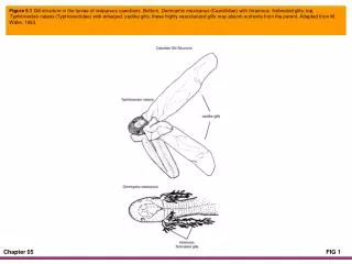
Figure 5.1 Gill structure in the larvae of viviparous caecilians. Bottom, Dermophis mexicanus (Caeciliidae) with triramous, fimbriated gills; top, Typhlonectes natans (Typhlonectidae) with enlarged, saclike gills; these highly vascularized gills may absorb nutrients from the parent. Adapted from M. Wake, 1993.
Figure 5.2 Females of the frog Leptobatrachium boringiae position themselves on the substrate under submerged rocks during asymmetrical inguinal aplexus while the male (top) pushes the eggs to the undersurface of the rock with his right hindleg. The end result is a mass of eggs that looks like a doughnut (insert). Adapted from Cheng and Fu, 2007, drawing by Z. Zheng.
Figure 5.3 Production of a foam nest by a paired male and female Leptodactylus knudseni. These large leptodactylids may deposit eggs in the same nest more than once. Tadpoles develop in the foam and are washed into a nearby pond if heavy rains occur. (W. Hödl, 1990)
Figure 5.4 Secretions from a male and female are whipped by rapid leg movements into a foam nest by the Brazilian leiuperid, Physalaemus ephippifer. At the same time, eggs are deposited and fertilized. The black circles represent the path of an egg as it is extruded from the female and pushed into the growing mound of foam; several hundred eggs will be deposited in a single nest. Adapted from Hödl, 1990.
Figure. 5.5 Larva of Leptodactylus labyrinthicus after eating eggs of other frog species inhabiting ponds in Goiás, Brazil. (A. Sebben)
Figure 5.6 From top to bottom: Mating ritual of Pipa parva. The pair somersaults in the water as eggs are released and fertilized; the male presses the eggs into the female's dorsum, where they embed in her skin. A female Pipa parva with freshly deposited eggs on her dorsum. Tadpoles emerging from pockets on the back of a female Pipa carvalhoi. (K.-H. Jungfer, 1996)
Figure 5.7 Direct-developing eggs of Pristimantis sp. In this species, eggs are deposited in leaf litter in a tropical forest. Note the well-developed back legs of the embryos. (J. P. Caldwell)
Figure 5.8 Frogs in the family Centrolenidae deposit their eggs on the undersides of leaves over moving water, where they develop into tiny tadpoles that drop into the stream. (J. P. Caldwell)
Figure 5.9 Left: Nest construction by a male Leptodactylus mystaceus; male calls from the depression to attract a female. Right: The male and a female produce a foam nest in which they deposit eggs. The nest is abandoned, and tadpoles are flooded from the nest when heavy rains occur. (J. P. Caldwell)
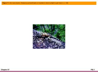
Figure 5.10 Synchronous hatching occurs when eggs of the Amazonian lizard, Plica plica, are disturbed. (L. J. Vitt)
Figure 5.11 Evolutionary events leading to vivparity and matrotrophy in vertebrates. Adapted from Blackburn, 2006.
Figure 5.12 Evolutionary events leading to viviparity and matrotrophy in squamates. CL refers to corpora lutea. Adapted from Blackburn, 2006.
Figure 5.13 Diagrammatic representation of the chorioallantoic placenta in Mabuya heathi. The placenta lies above the embryo and consists of hypertrophied uterine (maternal) and chorionic (fetal) tissue forming the placentome, the joint structure for nutrient transfer to the embryo, waste transfer to the female, and gaseous exchange. The interdigitating structures are the chorionic areolae, the site of transfer and exchange. Adapted from Blackburn and Vitt, 1992.
Figure 5.14 Generalized pattern of growth in embryos of the viviparous New World skink, Mabuya heathi. The embryo increases more than 74,000% of its freshly ovulated mass as the result of nutrient uptake from the female. Adapted from Blackburn and Vitt, 1992.
Figure 5.15 Clockwise from top left: Male Hyalinobatrachium valerioi attending three clutches of eggs of different ages; female of Stefania evansi brooding exposed eggs on its back; female Flectonotus fitzgeraldi brooding five eggs in dorsal pouches; an amplexing pair of Gastrotheca walkeri. Large, pale yellow eggs are expelled singly from the female's cloaca, fertilized by the male, and manipulated into the brooding pouch on the female's back. Photographs: H. valerioi, W. Hödl; all others, K.-H. Jungfer.
Figure 5.16 Female of the skink Plestiodon fasciatus attending her clutch of eggs. (L. J. Vitt)
Figure 5.17 Events leading to deposition of tadpoles of the dendrobatid frog Epipedobates tricolor. From top to bottom, amplexus, tadpole attendance, tadpole transport, and release of tadpoles in water. (K.-H. Jungfer)
Figure 5.18 Crocodylus palustris carrying newly hatched offspring to water. (J. W. Lang)
Figure 5.19 Froglets that have nearly completed their development on the back of a brooding female Stefania evansi. (K.-H. Jungfer)
Figure 5.20 Top: A female Anotheca spinosa feeding trophic eggs to her tadpoles. Begging behavior of the tadpoles may stimulate egg laying. Bottom: Trophic eggs consumed by a tadpole of Anotheca spinosa are visible through the transparent skin. (K.-H. Jungfer)
Figure 5.21 Top: Adult female of Leptodactylus ocellatus situated at the edge of her tadpole school. For perspective, the tadpoles just below the frog in the top panel are about 50 mm in total length. The female remains with the tadpole school and aggressively attacks intruders. Bottom: Tadpole school of Leptodactylus ocellatus from central Brazil. (J. P. Caldwell)
Figure 5.22 Adult female of the salamander Plethodon albagula attending her egg clutch. (S. E. Trauth)