Assessment of Orthotopic Xenograft Model: Pathological Findings in Tumor and Normal Tissues
10 likes | 119 Views
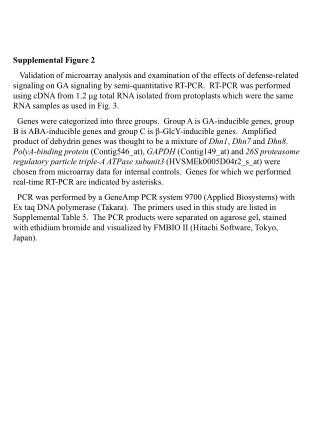
This study presents detailed histopathological images of an orthotopic xenograft model with viable and necrotic primary tumors, metastatic nodules in spleen/lung/bowel/diaphragm, and normal tissues post-treatment evaluation, revealing no cytotoxicity.

Assessment of Orthotopic Xenograft Model: Pathological Findings in Tumor and Normal Tissues
E N D
Presentation Transcript
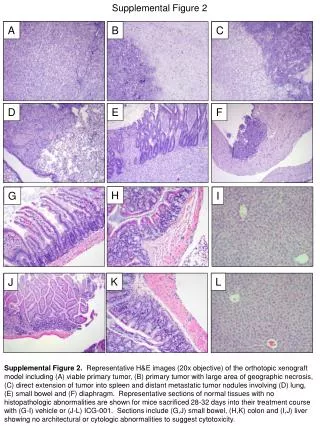
Supplemental Figure 2 A B C D E F H G I K L J Supplemental Figure 2. Representative H&E images (20x objective) of the orthotopic xenograft model including (A) viable primary tumor, (B) primary tumor with large area of geographic necrosis, (C) direct extension of tumor into spleen and distant metastatic tumor nodules involving (D) lung, (E) small bowel and (F) diaphragm. Representative sections of normal tissues with no histopathologic abnormalities are shown for mice sacrificed 28-32 days into their treatment course with (G-I) vehicle or (J-L) ICG-001. Sections include (G,J) small bowel, (H,K) colon and (I,J) liver showing no architectural or cytologic abnormalities to suggest cytotoxicity.