Download
1 / 20
210 likes | 481 Views
First and Early Second Trimester Diagnosis of Fetal Heart Disease. 성균관의대 소아과 삼성제일병원 진단방사선과 민 지 연. Benefits of Early Fetal ECHO. Early confirmation of normal cardiac anatomy Further testing, such as karyotyping Pharmacologic therapy Planned delivery Reduce early morbidity and mortality

E N D
First and Early Second Trimester Diagnosis of Fetal Heart Disease 성균관의대 소아과 삼성제일병원 진단방사선과 민 지 연
Benefits of Early Fetal ECHO • Early confirmation of normal cardiac anatomy • Further testing, such as karyotyping • Pharmacologic therapy • Planned delivery • Reduce early morbidity and mortality • Earlier and safer termination
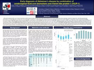
7.5 wks CRL; 1.5 cm Sonoembryology & Embryography • Heart beat; 6 wks (CRL 5.5 mm) • Septa, arterial & venous connection; after 8 wks • The mitral and tricuspid valve; 9-10 wks
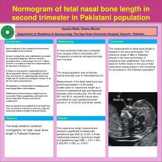
11.5 wks CRL; 5.8 cm sp sp Sonoembryology & Embryography • Aorta; end of 9 wks, larger than PA • AP position of IVS; prior to 11 wks • Brachiocephalic & carotid arteries; 12 wks
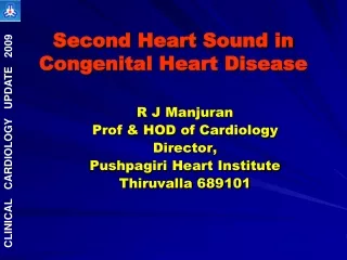
13.4 weeks CRL; 7 cm sp sp Sonoembryology & Embryography • LVOT, RVOT, Aortic arch with arterial duct; 13 wks • Complete four-chamber view; 13-14 wks (12 wks)
Transvaginal Ultrasonography TAS • 5-9 MHz • Transvaginal probe • 11-14 wks • For fetal heart • 11-14 weeks, TVS • 15-18 weeks, TVS=TAS • > 18 weeks, TAS TVS
Transvaginal Ultrasonography • Early diagnosis of CHD • Complex cardiac anomalies • Lesions with early chamber disproportion • Defects that are significant in size and/or severity • Limitation • Small size • Difficulties in spatial orientation • Limited range of imaging planes transabdominal sonography in 2nd trimester
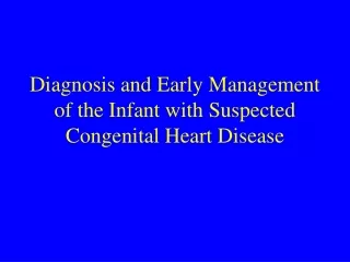
13 weeks CRL; 8 cm Situs solitus Dextrocardia Corrected TGA Functional single ventricle Pulmonary stenosis
sp R L 12 weeks, CRL; 6 cm NT; 4 mm Mesocardia Right Isomerism
R R sp ST sp L L 12.5 weeks TOF
Nuchal Translucency (NT) • Non-invasive firsttrimester US screening • Fluid collection at back of neck measured • 11-14 wks
Nuchal Translucency Screening • Combination of maternal age & NT; 80% detection rate for Down syndrome at 5% false positive rate • Prevalence higher than normal • Major cardiac defects • Diaphragmatic hernia • Exomphalos • Body stalk anomaly • Fetal akinesia
NT & Cardiac Defects • 29,154 fetuses with normal chromosomes • Prevalence of major cardiac defects < 95th percentile 0.8 > 95th percentile-3.4 mm 5.3 3.5-4.4 mm 28.9 4.5-5.4 mm 90.9 > 5.5 mm 195 Total 1.7 per 1000
NT, Possible Mechanism • Cardiac failure • not supported by range of CHD • no evidence in 2nd trimester of heart failure • BUT myocardial dysfunction in the 1st trimester?
NT & Ductus Venosus • Abnormal Doppler pattern (absence or reversal during atrial contraction); 90% of cases with chromosomal anomalies Normal Absent A Reversed A
NT & Ductus Venosus • Abnormal DV flow in euploid fetuses with increased NT helps to identify those with underlying CHD • 142 euploid fetuses with increased NT • Major CHD in 7/11 with abnormal flow • No CHD in 131 fetuses with normal flow
NT, Other Possible Mechanism • Venous congestion in head and neck • Failure of lymphatic drainage if impaired fetal movement • Abnormal or delayed development of lymphatic system • Altered composition of subcutaneous connective tissue
Embryonic & Fetal Heart Rate • Suspicion of CHD; cardiac decompensation • Spontaneous abortion
Precautions & Recommendations • Difficulties in pathological confirmation • More complex, more severe hemodynamic disturbance, frequent spontaneous miscarriage • Considerable experience • 13–15 weeks’ gestation • High-risk patients