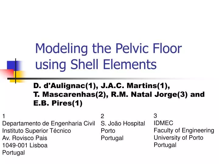

Modeling the Pelvic Floor using Shell Elements
E N D
Presentation Transcript
Modeling the Pelvic Floor using Shell Elements D. d'Aulignac(1), J.A.C. Martins(1), T. Mascarenhas(2), R.M. Natal Jorge(3) and E.B. Pires(1) 3IDMECFaculty of EngineeringUniversity of Porto Portugal 1Departamento de Engenharia Civil Instituto Superior Técnico Av. Rovisco Pais 1049-001 Lisboa Portugal 2S. João HospitalPortoPortugal
Finite Element Simulation Anatomy Plan Data Geometry
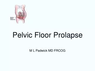
Why? • Understand working of the pelvic floor • Support of organs • Predict damages during childbirth • Stress incontinence • Prolapse • Uterine • Vaginal • Rectal
Pelvic Floor Illust. From Netter
MRI Images Sao Joao Hospital, Porto axial sagittal
Visualisation Segmented manually from MRI data. Julia R.Fielding et al. (Harvard medical school) Rasmussen et al (University of Illinois at Chicago)
Cadaver Measurements Janda et al 2003
Geometry Point Set NURB Surfaces (Rhino 3d) Polygon Mesh
Mesh Geometry top Frontal view of the mesh: 3068 triangles 1620 nodes side
3D Muscle Model Martins et al. 98 isotropic fibers volume
Muscle Model Deformation gradient Left Cauchy-Green tensor Right Cauchy-Green tensor
Muscle Model First Invariant Fibre strain Direction of fibres (deformed)
Plane Stress Since incompressibility is assumed Since normal stresses are zero the plane stress is given as
Passive Behaviour isotropic fibres Humphrey’s model for cardiac tissue
Passive Tests u lambda
Muscle Activation Sum of passive and active contributions
Total Stress isotropic total active fibers
Discussion • Large quantitative differences between different models • Oomens • Martins • Bosboom • Gielen • Kojic • Humphrey • Comparison with other models and experimental results is essential
Simulation 3068 triangular shell elements Non-linear simulation performed with ABAQUS UMAT routine decribing the material
The Future • Deformation of pelvic floor during childbirth • Damage, fracture of soft tissues • Prolapse of internal organs • Geometric models from MRI data • 8-node solid FE (de Sousa et al. 03)
Muscle Model Deformation gradient Left Cauchy-Green tensor Right Cauchy-Green tensor