Download
1 / 8
90 likes | 367 Views
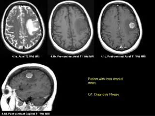
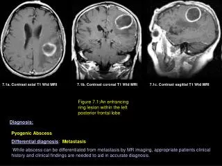
7.1a. Contrast axial T1 Wtd MRI. 7.1b. Contrast coronal T1 Wtd MRI. 7.1c. Contrast sagittal T1 Wtd MRI. Figure 7.1: An enhancing ring lesion within the left posterior frontal lobe. Diagnosis:. Pyogenic Abscess Differential diagnosis : Metastasis

E N D
7.1a. Contrast axial T1 Wtd MRI 7.1b. Contrast coronal T1 Wtd MRI 7.1c. Contrast sagittal T1 Wtd MRI Figure 7.1: An enhancing ring lesion within the left posterior frontal lobe Diagnosis: Pyogenic Abscess Differential diagnosis: Metastasis While abscess can be differentiated from metastasis by MR imaging, appropriate patients clinical history and clinical findings are needed to aid in accurate diagnosis.
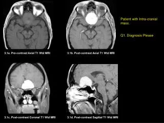
7.2a,b,c. Pre-Contrast axial T1 Wtd MRI 7.2d,e,f. Post-Contrast axial T1 Wtd MRI Figure 7.2: Multiple enhancing ring lesions in a immunocompromised patient Diagnosis: Fungal Abscesses Differential diagnosis: Multiple Metastasis While abscess can be differentiated from metastasis by MR imaging, appropriate patients clinical history and clinical findings are needed to aid in accurate diagnosis.
Fig 7.3a. T2-weighted images of the brain at just above the level of roof of the lateral ventricles of a young homosexual man. Findings: Diagnosis: Areas of T2-weighted hyperintensity within the deep paraventricular white matter (arrows). Human Immunodeficiency Virus (HIV) Encephalopathy A myelin-stained section of the brain specimen reveals severe diffuse myelin pallor (*). Horizontal section of the brain revealing abnormal left frontal lobe white matter from demyelinization (arrow).
Fig. 7.3b. T2-weighted image of the brain at the level of frontal horns of a young homosexual man Findings: Diagnosis: Foci of T2-weighted hyperintensity involving the peripheral white matter at or close to cortico-medullary junction (arrows) and left basal ganglia (arrow head). Progressive Multifocal Leukoencephalopathy
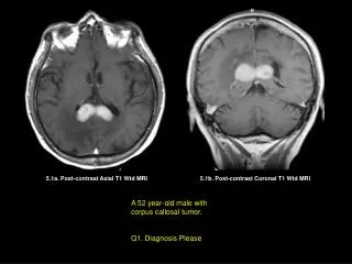
Fig 7.3c. Post-contrast axial CT image of the brain at the level of frontal horns/basal ganglia of a young homosexual man. Findings: Diagnosis: Enhancing ring lesion (arrow) within the left caudate nucleus. Primary CNS Lymphoma Coronal brain section reveals a tumor involving the left caudate nucleus (arrows). Photomicrographic show perivascular collections of lymphoma cells
Fig 7.3d. Post-contrast axial CT image of the brain at the level of the basal ganglia of a drug addict Findings: Diagnosis: Enhancing ring lesion (arrow) within the right caudate nucleus. CNS Toxoplasmosis Horizontal section of the right hemisphere reveals an irregular necrotic lesion in the right caudate nucleus
Fig. 7.3e. T1-weighted image of the brain at the level of frontal horns of a young homosexual man 7.3e. Pre-Contrast Axial T1 Wtd MRI Diagnosis: Findings: Focal area of T1 Wtd low signal intensity, capping the right frontal horn. Cytomegalo Virus Infection.
Fig 7.3f. Axial T2 Wtd image of the Brain. Diagnosis: Findings: Cryptococcal Meningitis Multiple small areas of increased T2 Wtd signal within both basal ganglia (arrows). Coronal brain section reveals multiple soap-bubble like lesions in the basal ganglia (arrows). Photo micrograph reveals encapsulated Cryptococcus neoformans with budding forms (arrow).