Download
1 / 70
700 likes | 732 Views
This informative text delves into the foundational discoveries of DNA structure by scientists like Griffith, Avery, Hershey, and Chargaff. It elucidates the intricate processes of DNA replication with detailed steps and mechanisms. Furthermore, it explores the genetic code, transcription, and translation, unveiling the flow of genetic information from DNA to protein synthesis in both prokaryotic and eukaryotic organisms. The concept of telomeres, proofreading, repair mechanisms, and the essential roles of various RNA types are also elucidated. This comprehensive guide provides a thorough understanding of DNA's significance and functionality in living organisms.

E N D
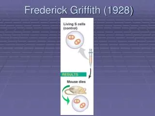
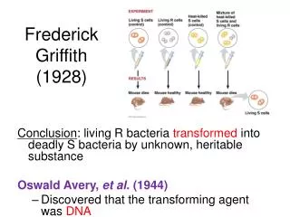
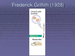
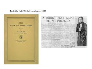
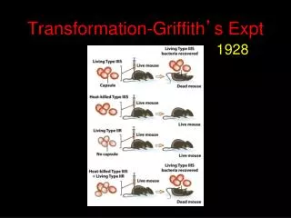
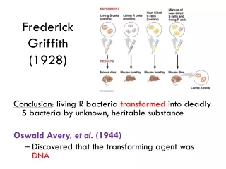
Frederick Griffith (1928) Conclusion: living R bacteria transformed into deadly S bacteria by unknown, heritable substance Oswald Avery, et al. (1944) • Discovered that the transforming agent was DNA
Hershey and Chase (1952) • Bacteriophages: virus that infects bacteria; composed of DNA and protein Protein = radiolabel S DNA = radiolabel P Conclusion: DNA entered infected bacteria DNA must be the genetic material!
Edwin Chargaff (1947) Chargaff’s Rules: • DNA composition varies between species • Ratios: • %A = %T and %G = %C
Structure of DNA Scientists: • Watson & Crick • Rosalind Franklin DNA = double helix • “Backbone” = sugar + phosphate • “Rungs” = nitrogenous bases
Structure of DNA Nitrogenous Bases • Adenine (A) • Guanine (G) • Thymine (T) • Cytosine (C) • Pairing: • purine + pyrimidine • A = T • G Ξ C purine pyrimidine
Structure of DNA Hydrogen bondsbetween base pairs of the two strands hold the molecule together like a zipper.
DNA Comparison Prokaryotic DNA Eukaryotic DNA Double-stranded Linear Usually 1+ chromosomes In nucleus DNA wrapped around histones (proteins) Forms chromatin • Double-stranded • Circular • One chromosome • In cytoplasm • No histones • Supercoiled DNA
Major Steps of Replication: • Helicase:unwinds DNA at origins of replication • Initiation proteins separate 2 strands forms replication bubble • Primase: puts down RNA primer to start replication • DNA polymerase III: adds complimentary bases to leading strand (new DNA is made 5’ 3’) • Lagging strand grows in 3’5’ direction by the addition of Okazaki fragments • DNA polymerase I: replaces RNA primers with DNA • DNA ligase: seals fragments together
Okazaki Fragments: Short segments of DNA that grow 5’3’ that are added onto the Lagging Strand DNA Ligase: seals together fragments
Proofreading and Repair • DNA polymerases proofread as bases added • Mismatch repair: special enzymes fix incorrect pairings • Nucleotide excision repair: • Nucleases cut damaged DNA • DNA polymerase and ligase fill in gaps
Nucleotide Excision Repair Errors: • Pairing errors: 1 in 100,000 nucleotides • Complete DNA: 1 in 10 billion nucleotides
Problem at the 5’ End • DNA polymerase only adds nucleotides to 3’ end • No way to complete 5’ ends of daughter strands • Over many replications, DNA strands will grow shorter and shorter
Telomeres: repeated units of short nucleotide sequences (TTAGGG) at ends of DNA • Telomeres “cap” ends of DNA to postpone erosion of genes at ends (TTAGGG) • Telomerase: enzyme that adds to telomeres • Eukaryotic germ cells, cancer cells Telomeres stained orange at the ends of mouse chromosomes
Flow of genetic information • Central Dogma: DNA RNA protein • Transcription: DNA RNA • Translation: RNA protein • Ribosome = site of translation • Gene Expression: process by which DNA directs the synthesis of proteins (or RNAs)
one gene = one polypeptide DNA RNA Nucleic acid composed of nucleotides Single-stranded Ribose=sugar Uracil Helper in steps from DNA to protein • Nucleic acid composed of nucleotides • Double-stranded • Deoxyribose=sugar • Thymine • Template for individual
RNA plays many roles in the cell • pre-mRNA=precursor to mRNA, newly transcribed and not edited • mRNA= the edited version; carries the code from DNA that specifies amino acids • tRNA= carries a specific amino acid to ribosome based on its anticodon to mRNA codon • rRNA= makes up 60% of the ribosome; site of protein synthesis • snRNA=small nuclear RNA; part of a spliceosome. Has structural and catalytic roles • RNAi= interference RNA; a regulatory molecule
The Genetic Code For each gene, one DNA strand is the template strand mRNA (5’ 3’) complementary to template mRNA triplets (codons) code for amino acids in polypeptide chain
The Genetic Code 64 different codon combinations Redundancy: 1+ codons code for each of 20 AAs Reading frame: groups of 3 must be read in correct groupings This code is universal: all life forms use the same code.
Transcription Transcription unit: stretch of DNA that codes for a polypeptide or RNA (eg. tRNA, rRNA) RNA polymerase: • Separates DNA strands and transcribes mRNA • mRNA elongates in 5’ 3’ direction • Uracil (U) replaces thymine (T) when pairing to adenine (A) • Attaches to promoter (start of gene) and stops at terminator (end of gene)
1. Initiation Eukaryotes: TATA box = DNA sequence (TATAAAA) upstream from promoter Transcription factors mustrecognize TATA box before RNA polymerase can bind to DNA promoter
2. Elongation • RNA polymerase adds RNA nucleotides to the 3’ end of the growing chain (A-U, G-C)
3. Termination RNA polymerase transcribes a terminatorsequence in DNA, then mRNA and polymerase detach. It is now called pre-mRNA for eukaryotes. Prokaryotes = mRNA ready for use
Additions to pre-mRNA: • 5’ cap(modified guanine) and 3’poly-A tail(50-520 A’s)are added • Help export from nucleus, protect from enzyme degradation, attach to ribosomes
RNA Splicing • Pre-mRNA has introns (noncoding sequences) and exons (codes for amino acids) • Splicing = introns cut out, exons joined together
RNA Splicing • small nuclear ribonucleoproteins = snRNPs • snRNP = snRNA + protein • Pronounced “snurps” • Recognize splice sites • snRNPs join with other proteins to form a spliceosome Spliceosomescatalyze the process of removing introns and joining exons Ribozyme = RNA acts as enzyme
Why have introns? • Some regulate gene activity • Alternative RNA Splicing: produce different combinations of exons • One gene can make more than one polypeptide! • 20,000 genes 100,000 polypeptides
Components of Translation • mRNA = message • tRNA= interpreter • Ribosome = site of translation
tRNA • Transcribed in nucleus • Specific to each amino acid • Transfer AA to ribosomes • Anticodon: pairs with complementary mRNA codon • Base-pairing rules between 3rd base of codon & anticodon are not as strict. This is called wobble.
Ribosomes • Ribosome = rRNA + proteins • made in nucleolus • 2 subunits 60s 40s
Ribosomes Active sites: • A site: holds AA to be added • P site: holds growing polypeptide chain • E site: exit site for tRNA
Translation:1. Initiation • Small subunit binds to start codon (AUG) on mRNA • tRNA carrying Met attaches to P site • Large subunit attaches
3.Termination • Stop codon reached and translation stops • Release factor binds to stop codon; polypeptide is released • Ribosomal subunits dissociate
Protein Folding • During synthesis, polypeptide chain coils and folds spontaneously • Chaperonin: protein that helps polypeptide fold correctly
Cell Cycle: life of a cell from its formation until it divides Functions of Cell Division: Reproduction, Growth and Tissue Renewal
Each chromosome must be duplicated before cell division Duplicated chromosome = 2 sister chromatids attached by a centromere
Gametes Somatic Cells Body cells diploid (2n): 2 of each type of chromosome Divide by mitosis Humans: 2n = 46 Sex cells (sperm/egg) Haploid (n): 1 of each type of chromosome Divide by meiosis Humans: n = 23
Phases of the Cell Cycle • The mitotic phase alternates with interphase: G1 S G2 mitosis cytokinesis • Interphase (90% of cell cycle) • G1 Phase: cell grows and carries out normal functions • S Phase: duplicates chromosomes • G2 Phase: prepares for cell division • M Phase (mitotic) • Mitosis: nucleus divides • Cytokinesis: cytoplasm divides
Mitosis 1. Prophase • Chromatin fibers condense and coil • Nucleoli disappear • Spindle (microtubules) begins to form • Centrosomes begin to move to opposite ends 2. Prometaphase • Nuclear envelope fragments • Microtubules invade nucleus • Kinetochores attach to microtubules
3. Metaphase • Chromosomes line up on metaphase plate at equator • Centrioles are at opposite poles (ends) 4. Anaphase (shortest phase) • Chromatids separate and pulled apart by motor proteins toward opposite ends of cell • Chromatids are called chromosomes now • Cell elongates
5. Telophase • Nuclear membrane re-forms around chromosomes • Chromosomes less condensed Cytokinesis • Cytoplasm of cell divided • Animal Cells: cleavage furrow • Plant Cells: cell plate forms
Cell Cycle Control System Checkpoint = control point where stop/go signals regulate the cell cycle
Major Checkpoints • G1 checkpoint (Most important!) • “Go” completes whole cell cycle • “Stop” cell enters nondividing state (G0 Phase) • Nerve, muscle cells stay at G0; liver cells called back from G0 • G2 checkpoint • M Phase checkpoint • Anaphase does not begin unless chromatids are properly attached to spindle at metaphase plate
Internal Regulatory Molecules • Kinases (cyclin-dependent kinase,Cdk): protein enzyme controls cell cycle; active when connected to cyclin • Cyclins: proteins which attach to kinases (Cdk) to activate them; levels fluctuate in the cell cycle 3. MPF: maturation-promoting factor; specific Cdk which allows cells to pass G2 and go to M phase
External Regulatory Factors • Growth Factor: proteins released by other cells to stimulate cell division • Density-Dependent Inhibition: crowded cells normally stop dividing; cell-surface protein binds to adjoining cell to inhibit growth • Anchorage Dependence: cells must be attached to another cell or ECM to divide