History of the Gram Stain Method: Understanding Gram-Positive and Gram-Negative Cells
210 likes | 262 Views
In 1884, Danish doctor Hans Christian Gram discovered the Gram staining method. Learn the differences between Gram-positive and Gram-negative cells, and the staining process. This technique is essential for bacterial identification. Dive into the world of microbiology!

History of the Gram Stain Method: Understanding Gram-Positive and Gram-Negative Cells
E N D
Presentation Transcript
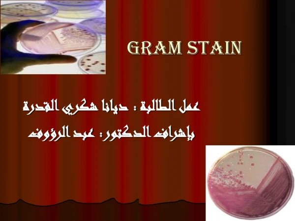
Gram stain عمل الطالبة : ديانا شكري القدرة بإشراف الدكتور: عبد الرؤوف
History of the Gram In 1884, Hans Christian Gram, a Danish doctor working in Berlin, accidentally stumbled on a method which still forms the basis for the identification of bacteria. While examining lung tissue from patients who had died of pneumonia The Gram staining method, named after the Danish bacteriologist who originally devised it
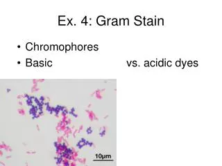
It is one of the most important staining techniques in microbiology.. It is almost always the first test performed for the identification of bacteria.. Gram developed a staining procedure which divided almost all bacteria into two large groups ..(Gram-positiveandGram-negative)
Gram positive and negative We first must to know what the Different between .. Gram positive and gram negative cells…
Gram positive cell The gram-positive cell envelope consists of a thick layer of peptidoglycan embedded with techoic acids and a plasma membrane comprised of phospholipids with integral membrane proteins traversing the bilayer..
Gram negative cell The gram-negative cell envelope consists of a thin layer of peptidoglycan surrounded by two phospholipid membranes, one interior and one exterior. Polysaccharide chains are bound to the phosphate heads of the outer membrane to form lipopolysaccharides..
Both the membranes contain integral membrane proteins. Place cursor over each membrane for ID…
Gram staining is based on the ability of bacteria cell wall to retaining the crystal violet dye during solvent treatment..
Gram stain procedure **Place a slide with a bacterial smear on a staining rack **stain the slide with crystal violet for 30-60 sec.
**Pour off the stain **Flood slide with Gram's iodine for 1-2 min **Pour off the iodine..
**Decolourize by washing the slide briefly with acetone (2-3 seconds).. **Wash slide thoroughly with water to remove the acetone – do not delay with this step..
**Flood slide with safranin counterstain for 30 sec.. **Wash with water.. **Blot excess water and dry in hand over bunsen flame..
What’s happen to gram negative cell when we stain it .. **The cells are flooded with crystal violet dye. ***The individual crystal violet ions penetrate the thin peptidoglycan layer of the cell as well as the plasma membrane, making their way through the matrix created by the crosslinking of polysaccharides and proteins within the peptidoglycan layer..
**Gram's iodine is added and penetrate the thin peptidoglycan layer of the cell. the iodide ions mix with the crystal violet dye.. **The crystal violet and iodide ions react, forming a crystal violet-iodine complex. This complex is insoluble in water and produces particles much larger than either the iodide ions or the crystal violet ions individually.
**A decolorizing solution, normally consisting of a mixture of ethyl alcohol and acetone, is added. **The mixture displaces water in the peptidoglycan layer, resulting in dehydration. This loss of water causes the thin peptidoglycan layer to shrink slightly, tightening the matrix created by the crosslinking of polysaccharides and proteins. The mixture also disrupts and dissolves the outer membrane, exposing the peptidoglycan layer to the environment.
Although the thin peptidoglycan layer of the gram-negative envelope is dehydrated, the crystal violet-iodide complex can escape through the large pores that remain. The complex is eventually washed away, leaving colorless, unstained cells, unlike gram-positive cells which appear purple at this step. ** The counterstain, normally safranin, is added.
What’s happen to gram positive cell when we stain it .. **The cells are flooded with crystal violet dye. Crystal violet is a water-soluble, basic dye **The individual crystal violet ions penetrate the thick peptidoglycan layer of the cell as well as the plasma membrane. **Gram's iodine solution is added **the iodide ions are also able to penetrate the thick peptidoglycan layer of the cell The crystal violet and iodide ions forming large complex ..
decolorizing solution, is added. It displaces water in the peptidoglycan layer, resulting in dehydration. This loss of water causes the thick peptidoglycan layer to shrink, tightening the matrix created by the crosslinking of polysaccharides and proteins. Because of its larger size, the crystal violet-iodine complex is blocked from moving easily through the thick layers of dehydrated peptidoglycan and exiting the bacterial cell.
**The counterstain, normally safranin, is added because of its small size of safranin is able to penetrate the dehydrated peptidoglycan layer **When viewed under a microscope, gram-positive cells appear purple due to the crystal violet-iodine complex retained inside