STAIN ING
STAIN ING. Department of Microbiology. Faculty of Veterinary Science. Chulalongkorn University. STAIN ING PROCEDURES;. Preparation of glass slide - remove grease & oil - water rinse & 95% alc - dry. 2. Preparation of smear - thin layer ( broth & solid medium) .

STAIN ING
E N D
Presentation Transcript
STAINING Department of Microbiology Faculty of Veterinary Science Chulalongkorn University
STAINING PROCEDURES; • Preparation of glass slide • - remove grease & oil • - water rinse & 95% alc • - dry 2. Preparation of smear - thin layer (broth & solid medium) 3. Heat fixation - protein coagulated & fixed to the slide surface
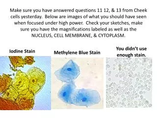
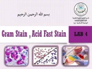
Study for : 1. SIMPLE STAIN 2. DIFFERENTIAL STAIN 2.1 GRAM STAIN 2.2 ACID-FAST STAIN (Ziehl-Neelsen’s method) 2.3 SPORE STAIN (Schaeffer-Fulton’s method)
Simple Stain MEDIA AND REAGENT EMPLOYED : Crystal violet 2% aq. solution.
METHOD : 1. Make thin film of organism smear, dry and fix with fire. 2. Flood with Crystal violet 1 min. 3. Rinse with tap water and drain or blot to dry. Vegetative cells are violet.
Differentialstaining Principle Differential staining : 1. Primary stain 2. Decolorizing agent 3. Counterstain
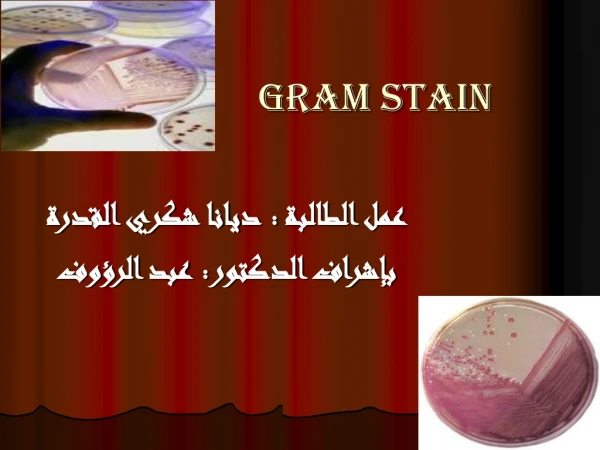
Gram Stain Dr. Christian Gram MEDIA AND REAGENT EMPLOYED : 1. Crystal violet 2% aq. solution. 2. Lugol’s solution. 3. Acetone+alcohol 4. Safranin 1% aq. solution.
Crystal violet Lugol soln insoluble complex + primary stain crystal violet - iodine (CV-I) complex + Mg CV-I Mg - CV - I complex
Gram + acetone Gram - small amount of lipid high concn of lipid minute cell wall pore large pores closed by dehydration of cell wall protein remain Mg - CV - I (blue) release CV-I complex safranin red or pink blue
METHOD : 1. Make a thin film of organism smear, dry and fix with fire. 2. Flood with Crystal violet 1 min. 3. Apply Lugol’s solution, remain for 1 min. 4. Rinse off the Lugol’s solution and decolourized with acetone, and wash by tap water.
METHOD : (continue) 5. Counterstain by Safranin for 30 sec to one min. 6. Wash with tap water. 7. Blot to dry and examine under oil immersion objective (x100). Gram’s positive are blueor deep blue. Gram’s negative are red or pink. Spores are no colour.
spore Vegetative cell
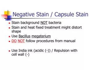
Acid - Fast Staining (Ziehl - Neelsen’s method) MEDIA AND REAGENT EMPLOYED : 1. Strong Carbol-fuchsin solution. 3. Acid alcohol solution. 2. 1% Malachite green solution.
Mycobacteria (Acid-fast bacteria) - thick waxy (lipodal cell wall). - penetration by stain extremely difficult. - cannot be readily removed by acid alcohol
Carbol fuchsin phenolic stain soluble in lipodal material retention of this stain penetration enhanced by heat to cytoplasm red tap water cool waxy cells substances to harden
non acid-fast bacteria Acid-alcohol acid-fast bacteria resistant lack cellular waxes retained red 1ry stain removed malachite green non - absorb absorb green red
METHOD : 1. Make organism smear, dry and fix with fire. 2. Flood the smear by strong carbol fuchsin, covered with filter paper or blotting paper and heat until the steam raise (but do not boil). 3. After 3-5 min. apply more heat until the steam raise again, do not let the stain dry on the smear.
METHOD : (continue) 4. About 5-7 min. after the first application of heat, wash the slide thoroughly by tap water. 5. Decolourize in acid alcohol until the tress of red disappeared from the smear. 6. Wash by tap water when decolorize completes.
METHOD : (continue) 7. Counterstain with Malachite green for 1 min. 8. Wash again by tap water, and stand on end to drain, do not blot to dry. Acid fast bacteria arered other bacteria are green
Spore Stain MEDIA AND REAGENT EMPLOYED : • Malachite green 5% aq. solution. • Tap water • 3. Safranin 0.5% aq. solution.
METHOD : 1. Make a thin smear of organism, dry and fix with fire. 2. Flood with 5% aq. malachite green and steam for 1-2 min. (cold stain - stand for 10 min). 3. Wash with tap water.
METHOD : 4. Counterstain with 0.5% aq. safranin for 15 seconds. 5. Rinse with water and drain or blot to dry. Vegetative cells of bacteria are red. Spores are green.
Bacterial culture broth Solid medium
Colony morphology Colony Size - 4mm, circular, entire Color - white Elevation - raised Texture - glisteining