Download

1 / 14
150 likes | 623 Views
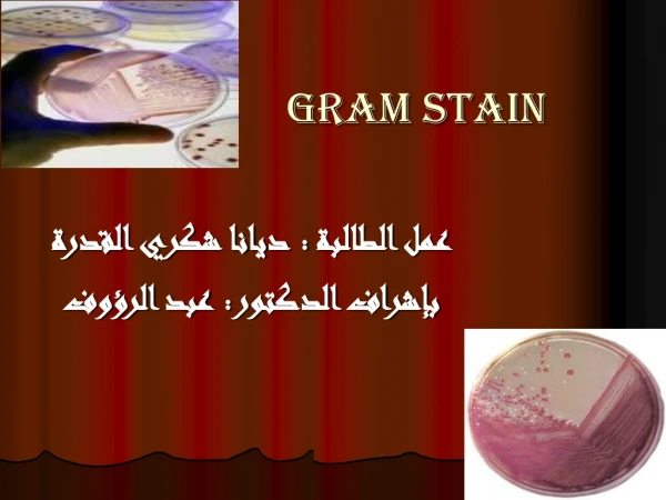
The Gram Stain. The Gram Stain. In the late 1800’s, Christian Gram observed that some genera of bacteria retained a dye-Iodine complex when rinsed with alcohol, while other genera were easily decolorized with alcohol and could be then visualized by a contrasting counterstain.

E N D
The Gram Stain In the late 1800’s, Christian Gram observed that some genera of bacteria retained a dye-Iodine complex when rinsed with alcohol, while other genera were easily decolorized with alcohol and could be then visualized by a contrasting counterstain. This staining procedure defines two bacterial groups: those which retain the primary dyes (“Positive by Gram’s Method” or “Gram-Positive”) and those which are easily decolorized (“Negative by Gram’s Method” or “Gram-Negative”). This is the starting point for bacterial identification procedures.
The Gram Stain The difference in dye retention is dependent on such physical properties as thickness, density, porosity, and integrity of the bacterial cell wall, as well as, to some extent, the chemical composition. Gram-Positive bacteria have thick, dense, relatively non-porous walls, while Gram-Negative bacteria have thin walls surrounded by lipid-rich membranes. See the Bacterial Morphology and Staining presentation for details. Some non-bacterial organisms with thick cell walls (e.g., some yeasts) also stain Gram-Positive. Gram-Positive bacteria which have lost wall integrity through aging or physical or chemical damage may stain Gram-Negative.
The Gram Stain Procedure • Step 1 - Prepare aSmear Suspend some of the material to be stained in a drop of water on a microscope slide, spread the drop to about the size of a nickel. Allow to air dry. Heat fix by gently warming above a flame or other heat source. Watch what happens to the “Bacteria” at each step “Bacteria”
The Gram Stain Procedure • Step 2 - Apply the Primary Stain Flood the Smear with Crystal Violet Allow to stand 30 sec to 1 min Rinse with water to remove excess stain
The Gram Stain Procedure • Step 3 - Apply the Fixing Agent Flood the Smear with Iodine solution Allow to stand 30 sec to 1 min
The Gram Stain Procedure • Step 4 - Rinse • Rinse with water to remove excess Iodine
The Gram Stain Procedure • Step 5 - Decolorize Drip 95% Alcohol across the slide about 5 sec The effluent should appear pale or clear
The Gram Stain Procedure • Step 6 - Rinse • Rinse with water to remove excess alcohol
The Gram Stain Procedure • Step 7 - Counterstain Flood the slide with Safranin solution Let stand 30 sec
The Gram Stain • Step 8 - Rinse,Dry and Observe Rinse with water to remove excess stain Blot dry Observe under Oil Immersion Gram-Positive Gram-Negative
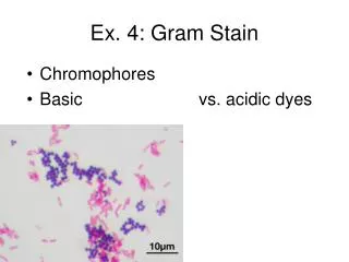
Examples of Gram Stains Gram Positive Rods and Cocci Gram Negative Rods and Cocci