Biomedical Instrumentation I
710 likes | 1.67k Views
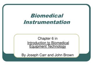
Biomedical Instrumentation I. Chapter 8 in Introduction to Biomedical Equipment Technology: Electrocardiography By Joseph Carr and John Brown. SA Node AV Node Bundle of His Bundle Branches Purkinjie Fibers. Schematic Representation of Electro-Conduction System.

Biomedical Instrumentation I
E N D
Presentation Transcript
Biomedical Instrumentation I Chapter 8 in Introduction to Biomedical Equipment Technology: Electrocardiography By Joseph Carr and John Brown
SA Node AV Node Bundle of His Bundle Branches Purkinjie Fibers Schematic Representation of Electro-Conduction System From Berne and Levy Physiology 3rd Edition Figure 23-25
Pathway of Electro-Conduction System of the calf heart starting at AV Node • AV Node • Bundle of His • Bundle Branches • Purkinjie Fibers From Berne and Levy Physiology 3rd Edition Figure 23-28
Components: P wave = Atrial Contraction QRS Complex = Ventricular Systole T Wave = Refractory Period Typical measurement from right arm to left arm Also see 1 mV Calibration pulse Electrocardiograph (ECG) Carr and Brown Figure 8-1
Different Segments of ECG P wave: the sequential activation (depolarization) of the right and left atria QRS complex: right and left ventricular depolarization (normally the ventricles are activated simultaneously) ST-T wave: ventricular repolarization U wave: origin for this wave is not clear - but probably represents "afterdepolarizations" in the ventricles PR interval: time interval from onset of atrial depolarization (P wave) to onset of ventricular depolarization (QRS complex) QRS duration: duration of ventricular muscle depolarization QT interval: duration of ventricular depolarization and repolarization RR interval: duration of ventricular cardiac cycle (an indicator of ventricular rate) PP interval: duration of atrial cycle (an indicator or atrial rate
Typical LeadsRA = right armLA = Left armLL = left legRL = right legC = ChestDifferent leads result in different waveform shapes and amplitudes due to different view and are called leads
Cardiac Axis by different Leads Carr and Brown Figure 8-2
Types of Leads Bipolar Limb Leads: are those designated by Lead I, II, III which form Einthoven Triangle: • Lead I = LA connection to noninverting amp. input And RA connecting to inverting amp. Input • Lead II = LL connection to amp. Noninverting input RA connect to inverting input and LA shorted to RL • Lead III = LL connected to noninverting input LA connected to inverting input LL LL LL
Einthoven Triangle:Note potential difference for each lead of triangle Carr and Brown Figure 8-3
Each lead gives a slightly different representation of electrical activity of heart
Types of Leads Unipolar Limb Leads=augmented limb leads: leads that look at composite potential from 3 limbs simultaneously where signal from 2 limbs are summed in a resistor network and then applied to an inverting amplifier input and the remaining limb electrode is applied to the non-inverting input Lead aVR = RA connected to non-inverting input while LA and LL are summed at inverting input augmented (amplified) Voltage for Right arm (aVR) Lead aVL = LA connected to non-invertinginput while RA and LL are summed at inverting input augmented (amplified) Voltage for Left arm (aVL) Lead aVF = LL connected to non-inverting input while RA and LA are summed at inverting input augmented (amplified) Voltage for Foot (aVF) LL LL LL
Unipolar Chest Leads: measured with signals from certain specified locations on the chest applied to amplifiers non-inverting input while RA LA, and LL are summed in a resistor Wilson network at amplifier inverting inputs Types of Leads LL
Configuration used with Unipolar Chest Leads where RA LA and LL are summed in resistor network and this is sent to the inverting input of an amplifier Wilson’s Central Terminal
Normal ECG with RA, LA, LL connected Artrial Tachycardia with RA, LA, LL connected Ventricular Tachycardiawith RA, LA, LL connected
Variations in Chest Leads C with RA and LA connected C1 C2 C3
1st Degree block RA LA LL connected PR wave is prolonged >0.2 sec have a prolongation of delay between atrial and ventricle depolarization Normal
2nd Degree Block Intermittent failure of AV conduction, such that not every P wave is followed by QRS complex Normal
3rd Degree Block Complete failure of conduction between atria nd ventricles. Common cause is AMI (Acute Myocardial Infarction Normal
R Bundle Branch Block Widened QRS complex abnormalities in R S as well as T wave Q is not as affected because the left bundle branch initiates depolarization Normal
Other ECG Signals • Interdigital ECG: Signal taken between 2 fingers usually for home monitoring • Esophageal ECG: electrode placed in esophagus close to heart typically used to record atrial activity where P and R wave are used to determine position • Toilet Seat ECG: used to detect cardiac arrhythmias that can occur during defecation
ECG Pre-Amplifier • High Impedance input of bioelectric amplifier • Lead selector switch • 1mV calibration source • Means of protecting amplifier from high voltage discharge such as a defibrillator used on a patient • Amplifier will have instrumentation amplifier as well as isolation amplifier
Needed for safety! Want to isolate patient from high voltages and currents to prevent electric shock where there is specifically a barrier between passage of current from the power line to the patient. Can be done using light (photo emitter and photo detector) or a transformer (set of inductors that are used in a step up / step down configuration) Isolation Amplifier
Common Mode Rejection • Until now we assumed Amplifiers were ideal such that the signal into each terminal would completely cancel lead to complete common mode rejection • However with practical Op Amp there is not perfect cancellation thus you are interested in what common mode rejection is.
Simplistic Example of ECG Circuit Would like to analyze what type of common mode voltage (CMV) can be derived
Common Mode Voltage (CMV) • If 2 inputs are hooked together into a differential amplifier driven by a common source with respect to ground the common mode voltage should be the same and the ideal output should be zero however practically you will see a voltage. • CMV is composed of 2 parts: • DC electrode offset potential • 60Hz AC induced interference caused by magnetic and electric fields from power lines and transformers • This noise is a current from in signal, common and ground wires • Capacitively coupled into circuit • (Other markets that work at 220-240 V will experience 50Hz noise)
Analysis to reduce noise in ECG • Common Mode Rejection: • Instrumentation amplifier (EX. INA128) using a differential amplifier which will cancel much of the 60 Hz and common DC offset currents to each input • If each signal is carrying similar noise then the some of the noise will subtract out with a differential amplifier
Right leg driver circuit is used in a feedback configuration to reduce 60 Hz noise and drive noise on patient to a lower level. Analysis to reduce noise in ECG
Thus Vn is reduced by Gain G1 Note Book forgot V in equation 5-35 Use of Feedback to reduce Noise Vn = Noise Vin V1G1 V1 V2 Vo V2G2 Σ G1 Σ G2 B Vo Β + + + Derivation:
Analysis to reduce noise in ECG • Isolation Amplifier also will attenuate noise • Shielding of cables further reduce noise
Review of Five ways to reduce Noise in ECG • Common Mode Rejection (differential Amplifier) • Right Leg Drive (feedback loop to decrease noise) • Shielding of wires • Isolation amplifier • Notch filter to reduce 60 Hz noise
How to overcome offset voltage Instrumentation Amplifier Gain (A1,A2,A3) = Non-Inverting Amplifier A4
Problems of offset voltage and how to correct • If you had 300 mV of DC offset sent through two gains of 10 and then 50 you would have an offset of (300mV)(10)(50) = 150V thus you would saturate your amplifiers and not see any of your signal • 3V offset after first set of noninverting amplifiers goes through differential amplifier A3 which reduces the offset voltage.
Other Corrections for Offset • Feedback circuit where output of A4 goes through HPF of A5 so only responses larger than cutoff frequency pass through thus the DC offset is attenuated R and C should be switched because this is really a LPF
Affect of High Pass Filter of A5 • Feedback through HPF has a time constant of RC • 3 Modes: • Diagnostic Mode (most time) where RC = 1x10-6F*3.2x106Ώ = 3.2 sec Cutoff Freq = 1/(2πRC) = 0.05Hz • Monitor Mode (medium time) where RC = 1x10-6F*318x103Ώ = 0.318 sec Cutoff Freq = 1/(2πRC) = 0.5Hz • Quick Restore (least time) where RC = 1x10-6F*80x103Ώ = 0.08 sec Cutoff Freq = 1/(2πRC) = 2Hz With Feedback the DC offset is eliminated and thus can have a gain of 50 on the 2nd Non-inverting Amplifier Stage without Saturating the Circuit Drawn Incorrectly R and C should be switched
High Pass Active Filters Attenuates High frequency where cutoff frequency is 1/(2) =1/ 2RiCi Rf Ci Ri - A Vinput + Voutput Ri Ci Voutput Rf Vinput 0 Ii IRf When frequencies (w) is small gain is reduced Gain (1Hz, Ci =1mF) When frequencies (w) is large gain ~ -Rf/Ri Gain (1MHz, Ci=1mF)
Low Pass Active Filters = Integrator Attenuates High frequency where cutoff frequency is 1/2=1/2RfCf Cf Rf Ri - A Vinput + Voutput Cf ICf Ri Rf Voutput Vinput 0 Ii IRf When frequencies (w) are high gain is reduced Gain (1M Hz, C=1mF) When frequencies (w) are low gain ~ -Rf/Ri Gain (0 Hz, C=1mF)
Defribillator • A Defribillator = a high voltage electrical heart stimulator used to resuscitate heart attack victims • When a physician applies this high voltage the high voltages and currents can cause damage to medical equipment BUT physician still needs to view ECG of the patient • How do you protect your medical equipment from excessively voltages and currents?
Glow Lamps are pair of electrodes mounted in a glass envelope in a atmosphere of lower pressure neon gain or a mix of inert gases Typically impedance across electrodes is high but if voltage across electrodes exceeds ionization potential of gas then impedance drops so you create a short to ground so vast majority of current goes safely to ground and avoids your amplifiers Protection Devices in ECGs: Glow Lamps
Diode: device that conducts electricity in one direction only Zener Diode: “Turns-On” when a minimum voltage is reached so in this configuration if a large voltage is applied (ie defibrillator) the zener diode will allow current to flow and shunts it to grounds thus current goes to ground and not to the amplifiers Protection Devices in ECGs: Zener Diodes
Diode: device that conducts electricity in one direction only Diode acts as a resistor as long as current level remains below limiting point. It current rises above the limit, the resistance will change and the current will become clamped Can also use a varistor (variable resistor) which functions like a surge protector that clips spikes in voltages Protection Devices in ECGs: Current-Limiting Diodes
Types of Defibrallitor Damage • Defibrillator is 6X greater than normal working voltage so damage will eventually occur • Two forms of Damage: • Both Amplifier inputs are blown thus readout is a flat line • One amplifier input is blown so the ECG appears distorted • Cause is from zener diodes becoming open or from glow lamps becoming defective from an air leak, or recombination or absorption of gases • Recommended that lamps are changed every 1-2 years or sooner if ECG is in Emergency Room
Sometime a high voltage transient is applied to the patient (defibrillator) which cause magnitudes much greater than biopotential signal (ECG) which saturates the amplifier Once the voltage transient signal is removed the ECG signal takes time to recover Effect of Voltage Transient on ECG
Example of bandwidth and magnitude of various biopotentials ECG is approximately 1 mV and spans from DC to 500 Hz Book assumes Diagnostic mode is 0.05 Hz to 100 Hz
Electro-Surgery Unit (ESU) Filtering • While a surgeon is conducting surgery he/she will want to see their patient’s ECG • ESU can introduce frequencies into the ECG of 100KHz to 100 MHz and with magnitudes up to kVolts which can distort the ECG • ESU introduces: • DC offsets • Obscures the signal • ESU needs to be of diagnostic quality thus you must eliminate higher frequencies which are noise
Correct for high frequency noise using LPF so ECG can function with ESU
RC Filters • Low Pass Filters will pass frequencies lower than cutoff frequency of FH =1/2RC Vs FH Frequency Vs FL • High Pass Filters will pass frequencies greater than cutoff frequency of FL =1/2RC