Download
1 / 31
320 likes | 364 Views
Explore the fascinating physiology and structure of the suprarenal (adrenal) glands, including the cortex and medulla. Learn about the functions of chromaffin cells, sympathetic nerve cells, and the intricate blood supply to these vital endocrine organs.

E N D
Endocrine Glands II Adrenal glands Prof Menna Abdel-Dayem
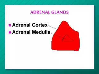
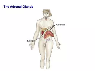
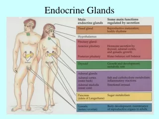
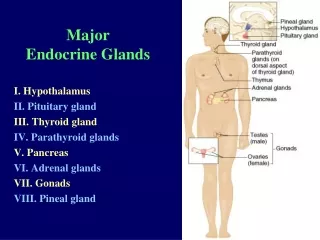
Two endocrine glands in contact with upper border of both kidneys. Derived from 2 embryonic sources: the mesoderm gives rise to adrenal cortex & neuroectoderm gives rise to adrenal medulla. Suprarenal (Adrenal) Glands
Stroma: Capsule, trabeculae and reticular framework Parenchyma: Cortex Medulla Suprarenal (Adrenal) Glands
Stroma: Capsule: thick, covered with adipose C.T. Trabeculae: thin, descend from the capsule to divide the cortex into compartments. Reticular framework Suprarenal (Adrenal) Glands
1. Chromaffin cells:– Modified sympathetic neurons that have lost their processes and became secretory cells.– Polyhedral cells arranged in branching cords separated by blood sinusoids.– Have basophilic cytoplasm rich in fine granules that stain brown with chromium salts & central rounded pale nuclei. Adrenal Medulla
1. Chromaffin cells: EM: • Cytoplasm shows mitochondria, well developed Golgi apparatus, rER & membrane-bound secretory granules. • There are 2 types of cells at ultrastructural level: a) Adrenalin secreting cells: with less dense & homogeneous granules, i.e. the contents fill the granules. b) Noradrenalin secreting cells: with more dense granules and peripheral clear halo beneath the membrane. The secretory granules also contain opiate-like peptides = enkephalins. Adrenal Medulla
Adrenal Medulla 2. Sympathetic nerve cells:- Stellate nerve cells scattered between chromaffin cells to stimulate their secretory activity.Innervated by preganglionic sympathetic nerve fibers.
Functions of the Adrenal Medulla • Secrete adrenalin (epinephrine) & noradrenalin (norepinephrine) in response to stress (fight, flight & fright). • During normal activity, the medulla continuously secretes small amounts of the 2 hormones.
Migrated sympathetic ganglion:1- Innervation ……….2- Embryologic origin …………3- contain …………
Paraganglia Masses of chromaffin cells scattered in close association with the sympathetic ganglia. They develop from neuroectoderm (like sympathetic ganglia). Give +ve chromaffin reaction. Function: secrete adrenalin & noradrenalin and enkephalins
Three suprarenal arteries (superior, middle & inferior) pierce the capsule → 3 groups of arterioles (capsular, cortical & medullary).The cortical capillaries drain into veins of medulla which join to form suprarenal vein that leave the gland Blood Supply