Download
1 / 32
330 likes | 528 Views
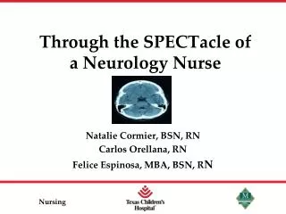
Through the SPECTacle of a Neurology Nurse. Natalie Cormier, BSN, RN Carlos Orellana, RN Felice Espinosa, MBA, BSN, R N. Objectives. Describe SPECT Scan and its purpose. Describe the role of a SPECT Nurse including safe administration of Neurolyte. Describe the characteristics of an EMU.

E N D
Through the SPECTacle of a Neurology Nurse Natalie Cormier, BSN, RN Carlos Orellana, RN Felice Espinosa, MBA, BSN, RN
Objectives • Describe SPECT Scan and its purpose. • Describe the role of a SPECT Nurse including safe administration of Neurolyte. • Describe the characteristics of an EMU.
Introduction What is SPECT? SPECT (Single Photon Emission Computed Tomography) imaging is a nuclear medicine study. It utilizes a radiopharmaceutical to create images of actual brain function by identifying blood flow patterns throughout the brain. Stewart, C. (2010). Single Photon Emission Computed Tomography (SPECT scan) basic level. Mayfield Clinic and Spine Institute. Retrieved from http://www.mayfieldclinic.com/PDF/PE-SPECT.pdf
Introduction • Brain perfusion SPECT is most commonly performed to aid in identification of epileptogenic focus in patients with medically refractory epilepsy (usually partial complex seizures) in whom surgical treatment is being considered. • It shows “hotspot” • In comparison to X-rays that display the internal structures of your body in 2-D image format, SPECT Scan generates 3-D images that display the actual working of the organs. • For example, a SPECT scan can display the movement of blood to your heart or areas inside your brain that may be more or less active. SPECT Scan. (2012). Mayo Foundation for Medical Education and Research (MFMER). Retrieved from http://www.mayoclinic.com/health/spect-scan/MY00233/DSECTION=risks
Introduction • The radioisotopes typically used in a SPECT scan to identify blood flow are: • iodine-123, • technetium-99m, • xenon-133, • thallium-201, • fluorine-18 • These radioactive forms of natural elements are safe and will pass safely through your body. The most commonly used isotope at Texas Children’s Hospital is technetium. • During a seizure, blood flow is highest at the point in the brain where the seizure starts. Stewart, C. (2010). Single Photon Emission Computed Tomography (SPECT scan) basic level. Mayfield Clinic and Spine Institute. Retrieved from http://www.mayfieldclinic.com/PDF/PE-SPECT.pdf
RISKS Risks Involved: • Pain, swelling or bleeding at the specific spot on your arm where the needle was inserted. • Although rare, allergic reaction may occur from the radioactive tracer. Radiation Risks: • During a SPECT scan, the least possible amount of radiation is used. • Radiation levels during SPECT scans are similar to what you may be exposed to over a period of one year in your surroundings. SPECT Scan. (2012). Mayo Foundation for Medical Education and Research (MFMER). Retrieved from http://www.mayoclinic.com/health/spect-scan/MY00233/DSECTION=risks
SPECT Imaging SPECT Scan Brain Images (2012). Retrieved from http://www.colin-studholme.net/research/ipag/ mrspect3.html.
Indication for SPECT scan Stewart, C. (2010). Single Photon Emission Computed Tomography (SPECT scan) basic level. Mayfield Clinic and Spine Institute. Retrieved from http://www.mayfieldclinic.com/PDF/PE-SPECT.pdf
NS FLUSH System Setup TRANSDUCER BASE PLATE RADIOIOSOTOPENEUROLYTE SYRINGE with lead syringe cover 3-WAY STOPCOCK TUBING
Procedure A peripheral IV is placed in a preferred site at the time of admission for the nurse to inject the radioactive tracer on the day of the SPECT scan The SPECT nurse and Child Life explain in developmentally appropriate ways about the SPECT scan prior to administration of the tracer.
Preparation for SPECT Procedure Our Radioactive Material Sign is placed on the patient’s door prior to the scan so that others will be aware of the test in progress. This also prevents any interruptions while the scan is in progress (i.e., the patient only seizes when she wakes up from a nap)
SPECT Procedure Nuclear Medicine calls the RN when the tracer is ready Nurse goes to Nuclear Medicine to pick up the lead box (above) with the tracer in a lead lining, RN Injection Protocol chart, and patient sticker
What to expect during a SPECT Scan? Usually, patient is made NPO if being sedated prior to SPECT Scan. SPECT Scan involves a two-step procedure: • Injection of radioisotope through the IV • Scanning of the brain Ictal SPECT • Radioactive material is administered during the seizure Interictal SPECT • Radioactive material is administered NOT during the seizure Pediatric Epilepsy Surgery Program. (2012). Single Proton Emission Computed Tomography (SPECT). Children’s Hospital of Pittsburg of UPMC. Retrieved from http://www.chp.edu/CHP/single+photon+emission+computed+tomography+(spect)
SPECT SCANs Picture courtesy of Child Life at Texas Chidren’s Hospital (with Parent Permission)
SPECT SCANs Picture courtesy of Child Life at Texas Chidren’s Hospital (with Parent Permission)
SPECT SCANs Picture courtesy of Child Life at Texas Chidren’s Hospital (with Parent Permission)
Nursing Implications - Training Picture courtesy of Child Life at Texas Chidren’s Hospital (with Parent Permission)
Nursing Implications - Education • Inform the patient about radioactive hazard and the procedure utilizing Child Life and their preparation book. • Address concerns and reassure the patient that the Radionuclide poses no radioactive hazard. • Reinforce importance of remaining still throughout the procedure because movement produces unreliable result. • There are no food, fluid, or medication restrictions unless by medical direction.
What you need to know after the test • A few hours after the SPECT scan, most of the radioactive substance is • flushed out by the body through the urine. • Patient needs to increase fluid intake to aid in removing the radioactive • substance. • Whatever is left behind is broken down by your body in one or two • days • SPECT Scan. (2011). Mayo Foundation for Medical Education and Research (MFMER). • Retrieved from http://www.mayoclinic.com/health/spect-scan/MY00233/DSECTION=risks After the SPECT Scan
Epilepsy Monitoring Unit (EMU) Texas Children’s Hospital EMU is considered level IV center among children's’ hospitals due to several factors, such as facilities, great cases, certain surgeries, EMG, SPECT Scans, etc. Goals: • Collaboration with key stakeholders regarding expansion • TCH Executives - EMU and Nursing Leaders/Team - Project Coordinator • Epileptologists - IS • Vendor
Epilepsy Monitoring Unit (EMU) Goals (cont) • Expansion of the EMU to 12-beds with state-of-the-art monitoring capabilities • Increase the volume of SPECTs, GRIDs, and possibly conduct a research study with bed expansions • Streamline admission process to the EMU and start VEEG study within a reasonable time • Maintain positive working relationship between EMU staff and nursing • Implementation of a dedicated RN EMU Team
The EMU Team - RNs • Consists of 11 RNs from days and 8 RNs from nights, 6 of which are SPECT nurses • Only team members will be assigned to EMU beds • Training for the team includes: • In-class training by Nuclear Medicine • Hands-on training to perform SPECT in Nuclear Medicine • In-class in service in regards to identifying types of seizures by EMU Faculty (Epileptologists) • Only a SPECT RN can administer isotope
Collaboration Between Nursing and EMU Staff • Nursing daily huddle in the EMU • Weekly meeting • Increase communication in regards to patient seizure activity between nursing and EMU techs • Meet and greet with the new EMU staff • Increase communication in regards to patient admission • Utilize EMU admission algorithm (next slide) • Green Belt Project
Green Belt Project IMPROVE Identify, prioritize, and pilot best solution(s) CONTROL Implement controls to sustain success Train all necessary personnel Audit periodically to validate • DEFINE • Identify the problem in the EMU • MEASURE • Document the current process • Quantify baseline metrics • Identify quick wins • ANALYZE • Identify potential causes
EMU – What’s in the horizon? • Expansion to a 6-bed EMU is underway • Expand to a 12-bed unit is the ultimate goal • Continue the Green Belt project • Maintain collaboration with EMU and Epileptologists • Review other initiatives to deliver quality patient care • Improve patient satisfaction • Maintain Level IV EMU center
References Mullen, J. (2012). EMU Admission Process Flowchart. Texas Children’s Hospital. Pediatric Epilepsy Surgery Program. (2012). Single Proton Emission Computed Tomography (SPECT). Children’s Hospital of Pittsburg of UPMC. Retrieved from http://www.chp.edu/CHP/single+photon+emission+computed+tomography+(spect) Scanning Images (2011). Child Life Department. Texas Children’s Hospital . SPECT Scan. (2011). Mayo Foundation for Medical Education and Research (MFMER). Retrieved from http://www.mayoclinic.com/health/spect-scan/MY00233/DSECTION=risks SPECT Scan Brain Images (2012). Retrieved from http://www.colin-studholme.net/research/ipag/ mrspect3.html. Stewart, C. (2010). Single Photon Emission Computed Tomography (SPECT scan) basic level. Mayfield Clinic and Spine Institute. Retrieved from http://www.mayfieldclinic.com/PDF/PE-SPECT.pdf Texas Children’s Hospital (2011-2012). Epilepsy Monitoring Unit.