Download
1 / 44
440 likes | 468 Views
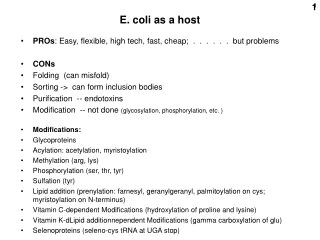
1. E. coli as a host. PROs : Easy, flexible, high tech, fast, cheap; . . . . . . but problems CONs Folding (can misfold) Sorting -> can form inclusion bodies Purification -- endotoxins Modification -- not done (glycosylation, phosphorylation, etc. ) Modifications:

E N D
1 E. coli as a host • PROs: Easy, flexible, high tech, fast, cheap; . . . . . . but problems • CONs • Folding (can misfold) • Sorting -> can form inclusion bodies • Purification -- endotoxins • Modification -- not done (glycosylation, phosphorylation, etc. ) • Modifications: • Glycoproteins • Acylation: acetylation, myristoylation • Methylation (arg, lys) • Phosphorylation (ser, thr, tyr) • Sulfation (tyr) • Lipid addition (prenylation: farnesyl, geranylgeranyl, palmitoylation on cys; myristoylation on N-terminus) • Vitamin C-dependent Modifications (hydroxylation of proline and lysine) • Vitamin K-dLipid additionnependent Modifications (gamma carboxylation of glu) • Selenoproteins (seleno-cys tRNA at UGA stop)
2 • Some alternative hosts • Yeasts (Saccharomyces , Pichia) • Insect cells with baculovirus vectors • Mammalian cells in culture (later) • Whole organisms (mice, goats, corn) • In vitro (cell-free), for analysis only(good for radiolabeled proteins
3 GAPDterm LEU2 GAPDprom Ampr oriE Yeast Expression Vector (example) Saccharomyces cerevisiae(baker’s yeast) 2 micron seq: yeast ori oriE = bacterial ori Ampr = bacterial selection LEU2, e.g. = Leu biosynthesisfor yeast selection 2 micron plasmid Complementation of an auxotrophy can be used instead of drug-resistance Your favorite gene(Yfg) Auxotrophy = state of a mutant in a biosynthetic pathway resulting in a requirement for a nutrient GAPD = the enzyme glyceraldehyde-3 phosphate dehydrogenase
Vector DNA t p gfY Genomic DNA Genomic DNA HIS4 mutation- Yeast - genomic integration via homologous recombination HIS4 Homologous recombination t p Yfg FunctionalHIS4 gene DefectiveHIS4 gene
HIS4 Vector DNA AOX1t Yfg AOX1p 3’AOX1 Genomic DNA AOX1 gene (~ 30% of total protein) Genomic DNA Yfg 3’AOX1 AOX1p AOX1t HIS4 Double recombination Yeast (integration in Pichia pastoris) P. pastoris-tight control-methanol induced (AOX1)-large scale production (gram quantities) His- host Alcohol oxidase gene
Protein-protein interactions Yeast 2-hybrid system Yeast 3-hybrid and 1 hybrid systems Co-immunoprecipitation Pull-downs Far western blots Biacore (surface plasmon resonance, SPR) Fragment complementation
Yeast 2-hybrid system: To discover proteins that interact with each other, or To test for interaction based on a hypothesis for a specific protein. Positive control: (bait) ? Y = e.g., a candidate protein being tested for possible interaction with X Or: Y = e.g., a cDNA library used to discover a protein that interacts with X ? (prey) BD =(DNA) binding domain AD =activation domain UAS =upstream activating sequence http://www.mblab.gla.ac.uk/~maria/Y2H/Y2H.html
No interaction between X and Y: no reporter expression Yes, interaction between X and Y: reporter protein is expressed Y = e.g., a cDNA library used to discover a protein that interacts with X Recover the Y sequence from reporter+ colonies by PCR to idenify protein Y
Fusion library Bait protein is the known target proteinfor whom partners are sought =“prey” Two different assays help, as there are often many false positives. BD= DNA binding domain; TA = transactiavting domain http://www.mblab.gla.ac.uk/~maria/Y2H/Y2H.html
3-HYBRID: select for proteins domains that bind a particular RNA sequence Use a known tight protein-RNA interaction (e.g., from RNA phage MS2) Prey Bait Prey could be proteins from a cDNA library
Yeast one-hybrid: Insert a DNA sequence upstream of the selectable or reporter Transform with candidate DNA-binding proteins (e.g., cDNA library) fused to an activator domain. Each T = one copy of a DNA target sequence
Indirect selection using a yeast 3-hybrid system:toward a more efficient glycosynthase enzyme Directed Evolution of a Glycosynthase via Chemical Complementation Hening Lin,† Haiyan Tao, and Virginia W. Cornish J. AM. CHEM. SOC. 2004, 126, 15051-15059 Turning a glycosidase into a glyco-synthase Glycosidase: Glucose-Glucose (e.g., maltose) + H2O 2 Glucose
Indirect selection using a yeast 3-hybrid system(one of the hybrid molecules here is a small molecule) e.g., from a mutated library of enzyme glycosynthase genes glucose Leu2 gene Leu2 gene Transform a yeast leucine auxotroph. Provide synthetic chimeric substrate molecules. Select in leucine-free medium. DHFR = dihydrofolate reductase GR = glucocorticoid receptor (trancription factor ) MTX = methotrexate (enzyme inhibitor of DHFR) DEX = dexamethasone, a glucocorticoid agonist, binds to GR AD = activation domain, DBD = DNA binding domain
Selection of improved cellulases via the yeast 2-hybrid system Survivors are enriched for cellulase genes that will cleave cellulose with greater efficiency (kcat / Km) Yeast cell Cellobiose (disaccharide) URA-3 (toxic) cellulase Directed Evolution of Cellulases via Chemical Complementation. P. Peralta-Yahya, B. T. Carter, H. Lin, H. Tao. V.W. Cornish. x x x x Library of cellulase mutant genes (one per cell)
URA-3 = gene for orotidine phosphate (OMP) decarboxylase Pathway to pyrimidine nucleotides: How does the URA-3 “suicide” system work? analog 5-fluoroorotic acid 5-fluoro-OMP URA-3 decarboxylation (pyr-4) 5-fluoro-UMP uridine kinase exogenousuridine thymidylate synthetase inhibition RNA death
Measuring protein-protein interactions in vitro X=one protein Y= another protein Pull-downs: Binding between defined purified proteins, at least one being purified. Tag each protein differently by making the appropriate cDNA clone. Examples: His6-X + HA-Y; Bind to nickel ion column via X, elute (his), Western with HA Ab for Y GST-X + HA-Y; Bind to glutathione ion column, elute (glutathione), Western with HA Ab His6-X + 35S-Y (made in vitro); Bind Ni column, elute (his), gel + autoradiography. No antibody needed. (HA = flu hemagglutinin) glutathione = gamma-glutamyl-cysteinyl-glycine.
Example of a result of a pull-down experiment Also identfy by MW (or mass spec) Total protein: no antibody/Western (stained with Coomassie Blue or silver stain) Antibody used in Western Compare pulled down fraction (eluted)with loaded. Loaded sample usually only a fraction.
Western blotting To detect the antibody use a secondary antibody against the primary antibody (e.g, goat anti-rabbit IgG). The secondary antibody is a commercial fusion protein with an enzyme activity (e.g., alkaline phosphatase). The enzyme activity is detected by its catalysis of a reaction producing a luminescent compound. * * http://www.bio.davidson.edu/courses/genomics/method/Westernblot.html
Detection of antibody binding in western blots Antibody to protein on membrane Alkaline phosphatase fusion Non-luminescent substrate-PO4 = Y Y Luminescent product + PO4= (chemiluminescence) Secondary antibody Detect by exposing to film Protein band on membrane
Far western blotting to detect specific protein-protein interactions. Use a specific purified protein as a probe instead of the primary antibody To detect the protein probe use an antibody against it. Then a secondary antibody against the first antibody, a fusion protein with an enzyme activity. The enzyme activity is detected by its catalysis of a reaction producing a luminescent compound. protein protein OR:Use a radioactively labeledprotein if interest and detect by autoradiography http://www.bio.davidson.edu/courses/genomics/method/Westernblot.html
Expression via in vitro transcription followed by in vitro translation T7 RNA polymerase binding site (17-21 nt) VECTOR cDNA ….ACCATGG….. Radioactively labeled protein 1. Transcription to mRNA via the T7 promoter + T7 polymerase 2. Add a translation system: rabbit reticulocyte lysate or wheat germ lysate Or: E. coli lysate (combined transcription + translation) All commerically available as kits Add ATP, GTP, tRNAs, amino acids, label(35S-met), May need to add RNase (Ca++-dependent) to remove endogenous mRNA In lysate NOTE: Protein is NOT at all pure (100s of lysate proteins present), just ~“radio-pure”
Surface plasmon resonance (SPR) Popular instrument is a Biacore The binding events are monitored in real-time and it is not necessary to label the interacting biomolecules. glass plate Reflection angle changes depending on the mass of the material on the surface. Binding increases this mass. Follow as a functrion of concentration Kd’s Or time : Measure on-time, off time; Kd = off-time/on-time http://home.hccnet.nl/ja.marquart/BasicSPR/BasicSpr01.htm
Expression in mammalian cells Lab examples: HEK293 Human embyonic kidney (high transfection efficiency) HeLa Human cervical carcinoma (historical, low RNase) CHO Chinese hamster ovary (hardy, diploid DNA content, mutants) Cos Monkey cells with SV40 replication proteins (-> high transgene copies) 3T3 Mouse or human exhibiting ~regulated (normal-like) growth + various others, many differentiated to different degrees, e.g.: BHK Baby hamster kidey HepG2 Human hepatoma GH3 Rat pituitary cells PC12 Mouse neuronal-like tumor cells MCF7 Human breast cancer HT1080 Human with near diploid karyotype IPS induced pluripotent stem cells and: Primary cells cultured with a limited lifetime. E.g., MEF = mouse embryonic fibroblasts, HDF = Human diploid fibroblasts Common in industry: NS1 Mabs Mouse plasma cell tumor cells Vero vaccines African greem monkey cells CHO Mabs, other therapeutic proteins Chinese hamster ovary cells PER6 Mabs, other therapeutic proteins Human retinal cells
Mammalian cell expression Generalized gene structure for mammalian expression: polyA site intron Mam.prom. 3’UTR cDNA gene 5’UTR Intron is optional but a good idea
SV40 LargeT Ag (Simian Virus 40) RSV LTR (Rous sarcoma virus) MMTV (steroid inducible) (Mouse mammary tumor virus) HSV TK (low expression) (Herpes simplex virus) Metallothionein (metal inducible, Cd++) CMV early (Cytomegalovirus) Engineered inducible / repressible:tet, ecdysone, glucocorticoid (tet = tetracycline) Popular mammalian cell promoters
Engineered regulated expression: Tetracycline-reponsive promoters Tet-OFF (add tet shut off) Tet-OFF VP16 tc’nact’n domain tetRdomain tTA = tet activator fusion protein: active No tet.Binds tet operator(if tet not also bound) Tet-OFF VP16 tc’nact’n domain tetRdomain Allosteric change in conformation Tetracycline (tet), or,better, doxicyclin (dox) not active tTA gene must be in cell (permanent transfection, integrated): polyA site CMV prom. tTA cDNA (Bujold et al.)
tetRdomain VP16 tc’nact’n domain not active little transcripton (2%?, bkgd) Doxicyclin present: polyA site MIN. CMV prom. your favorite gene polyA site polyA site your favorite gene your favorite gene No doxicyclin: VP16 tc’nact’n domain tetRdomain active Plenty of transcripton RNA po l MIN. CMV prom. Tet-OFF MIN. CMV prom. Mutliple tet operator elements
Tet-ON Tetracycline-reponsive promoters Tet-ON (add tet turn on gene tetRdomain VP16 tc’nact’n domain not active Different fusion protein: Does NOT bind tet operator(if tet not bound) tetRdomain VP16 tc’nact’n domain active Tetracycline (tet), or,better, doxicyclin (dox) polyA site Full CMV prom. tTA cDNA Must be in cell (permanent transfection, integrated): commercially available (293, CHO) or do-it-yourself
polyA site polyA site polyA site your favorite gene your favorite gene your favorite gene Tet-ON MIN. CMV prom. Mutliple tet operator elements tetRdomain VP16 tc’nact’n domain not active little transcription (bkgd.) Doxicyclin absent: MIN. CMV prom. Add dox: active tetRdomain VP16 tc’nact’n domain doxicyclin active Plenty of transcripton (> 50X) RNA pol II MIN. CMV prom.
Reporterenzyme Enzyme fragments themselves do not associate well enough to reconstitute an active enzyme F = reporter protein fragment SW Michnick web site: http://michnick.bcm.umontreal.ca/research/images/pca_general_en.gif
Clonal selection and in vivo quantitation of protein interactions with protein-fragment complementation assays, I. Remy and S.W. Michnick PNAS 96, 394–5399, 1999 DHFR fragments Rapamycin promotes the association of the 2 protein domains fMTX Fluorescein-MTX IN PURINE-FREE MEDIUM DHFR = dihydrofolate reductase DHF=dihydrofolate = FH2 THF=tetrahydrofolate = FH4 fMTX=fluorescent methotrexate FK506 = immunosuppressant drug FKBP = FK506 binding protein FRAP = FKBP–rapamycin binding proteinFRB= FKBP–rapamycin binding domain of FRAP
FK506 recruits FKBP to bind to calcineurin and inhibit its action as a specific phosphatase a phosphatase
Claim detection of 0.05 nM rapamycin ?? No. of CHO colonies [rapamycin]
Fluorescent methotrexate (fMTX) assay CHO cells (permanent transfection) cos cells (transient transfection) Leucine zipper protein fragments instead of rapamycin binding proteins (positive contro) Background association of FKBP and FRB without rapamycin (compare)
8-fold increase in fluorescence per cell No. of cells fluorescence-activated flow cytometer (FACS is this plus more) Log of fluorescence intensity Fluorescence intensity Measure affinity for a drug in vivo [rapamycin] Competition with a molecule that binds only one
Erythropoietin-erythropoietin receptor (dimer) interaction: Efficacy of a peptide mimetic Erytropoietin (EPO) receptor In vivo assay of drug effectiveness (EMP1) (inexpensive substitute for erythropoietin?) EMP1 = Erythropoietin mimetic peptide 1 Erythropoietin
FACS = Fluorescence-activated cell sorter Impart a charge on the recognized cell Less than one cell or particle per droplet. Thus the most that most droplets contain is one particle. Can be used purely anaytically without the sorting capability. Then called “flow cytometry”, or also called FACS anyway. Charged plates attract droplets containing a particle of the opposite charge Cells remain viable if treated with care.
Histogram-type display No fluorescence (background autofluorescence) No. of cells Red stained Usually a log scale Having this much fluorescence
Scatter plot display Analysis on 2 colors One cell Amount of greenfluorescence (log) You decide on the positions of of demarcations Say, want high reds but low greens: Instruct the FACS to deflect cells in this quadrant only. Collect and grow or analyze further. Amount of red fluorescence (log)
Beaming bead FACS analysis Analysis of beads representing the human genome using allele-specific hybridization probes and the FACS A. Flow cytometry data: 2-D plots where each point represents one particle. Then contour lines plotted around the point density. Here light “forward” scattering (irrespective of wavelength) is measured (FSC). Instrument can be set to reject data from 2-bead doublets that scatter light more. Both signals Red signal Neither signal Green signal B-D. Amplified beads hybridized to 2 probes, one specific to the S allele of a certain gene and one specific to the L allele. The beads carry the amplified PCR products corresponding to this region from 3 human individuals. The blue points come from microspheres that contained both types of PCR products from both alleles, despite the high dilution.