Download

1 / 25
250 likes | 638 Views
Stickler Syndrome Study at the National Institutes of Health Nazli McDonnell M.D., Ph.D. Laboratory of Clinical Investigation National Institute on Aging National Institutes of Health Baltimore, Maryland. Introduction.

E N D
Stickler Syndrome Study at the National Institutes of HealthNazli McDonnell M.D., Ph.D. Laboratory of Clinical Investigation National Institute on Aging National Institutes of Health Baltimore, Maryland
Introduction • Stickler Syndrome is an autosomal dominant hereditary disorder that effects the connective tissue • 1 in 10,000 individuals in North America (Hermann et al, 1975) are thought to have Stickler Syndrome • Stickler Syndrome may affect your eyes, your hearing, your bones and joints
Intro cont. • Known mutations that lead to Sticklers are found on the Col2A1, Col11A1 and the Col11A2 genes • They are genes code for the collagens that are found in your eyes, joints and bones • Mutations on these genes account for around 50% of the cases of Stickler Syndrome. Other causes for Sticklers syndrome are unknown at this time.
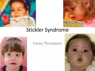
Manifestations • Stickler Syndrome effects many parts of the body. • In the eyes, Stickler syndrome may cause near-sightedness (myopia), vitreous degeneration, retinal detachments and retinal tearing, premature cataracts and glaucoma • People with Stickler Syndrome may be born with cleft palates and/or bifid uvulas
Manifestations cont. • The most noticeable manifestations of Stickler are the facial manifestations. People with Sticklers Syndrome often have a flattened facial profile, flattened and broadened nasal bridge and small chins. • Patients also have problems with high frequency hearing loss and hypermobile tympanic membranes
Diagnosis is often missed • Stickler syndrome is among the most common autosomal dominant connective tissue disorders but is often unrecognized and therefore not diagnosed by clinicians. • Ten percent of patients with isolated cleft palate and 12% with the Pierre-Robin sequence were found to have undiagnosed Stickler syndrome in one series [Kronwith et al., 1990; Sheffield et al., 1987] The actual incidence is higher.
Background • Collagens are the most common proteins in the extracellular matrix • Collagen Type II is abundant in the vitreous of the eye, the spinal column, cartridge, and inner ear • Type II is also present in many tissues during embryological development • Collagen XI is found in association with Collagen II.
The Collagens • Collagens consists of three polypeptide chains which are folded into a rod-like triple helical molecule • Each of the constituent chains of the triple helix are called alpha chains and are coiled in a left handed helix with three amino acids per turn. • The constituent amino acids are regularly arranged in the order Gly-X-Y such that glycine, which is the smallest of all amino acids, occupies the restricted space in which the three helical chains come together. • This arrangement is crucial for the stability of the macromolecule.
Collagen structure Side view shows: Primary structure = (X-Y-gly)n X,Y are often lysine or proline Top end view shows: Secondary structure is a collagen helix with pitch of 3.0 residues per turn
Collagens and Stickler Syndrome • Type 2 collagen is a homotrimer of three COL2A1 gene products, whereas type 11 collagen is a heterotrimer containing one each of the COL2A1, COL11A1, and COL11A2 gene products. • Both type 2 and 11 collagens are members of the fibrillar collagens, which are primarily found in cartilage, vitreous, and nucleus pulposus (soft, gelatinous central portion of an intervertebral disk).
The diagnostic criteria • Orofacial Abnormalities (2 points maximum) • (2 points) Cleft palate (open cleft, submucous cleft, or bifid uvula) (Major) • (1 point) Characteristic facies (malar hypoplasia, broad nasal bridge, • Micro/retrognathia) • Ocular Abnormalities (2 points maximum) • (2 points) Characteristic vitreous changes or retinal abnormalities (lattice degeneration, retinal holes, or retinal tear, retinal detachment) (Major) • Auditory Abnormalities (2 points maximum) • (2 points) High frequency sensorineural hearing loss (Major) • Age < 20: threshold 20 dB at 4-8 kHz • Age 20-40: threshold 30 dB at 4-8 kHz • Age > 40: threshold 40 dB at 4-8 kHz • (1 point) Hypermobile tympanic membranes • Skeletal Abnormalities (2 points maximum) • (1 point) History of femoral head failure • (slipped epiphysis or Legg-Perthes like disease) • (1 point) Radiographically demonstrated osteoarthritis before age 40 • (1 point) scoliosis, spondylolisthesis, or Scheuermann-like kyphotic deformity • Family History / Molecular Data • (1 point) Independently affected 1st degree relative in a pattern consistent with autosomal dominant inheritance or presence of COL2A1, COL11A1, or • COL11A2 mutation associated with Stickler syndrome • Diagnosis requires: 5 or more points total up to 9 points • At least one 2-point major manifestation • Absence of features suggestive of a skeletal dysplasia (e.g. stature <5% centile)
Genotype/Phenotype Correlations • Study involved 48 families suspected of having Stickler Syndrome • Consent was received from each individual to allow the NIA to collect data and genetic material from each patient • Patients underwent a detailed clinical genetics examination, dilated ophthalmology exam, audiology and otolaryngology evaluations
Materials and methods contd. • In most cases, radiographs of the spine and hip were available • The enrollment and tests took place before the development of the diagnostic criteria for Stickler syndrome. • Forty eight probands were selected from each family for molecular genetic analysis • Col2A1 and Col11A1 genes were amplified by polymerase chain reaction (PCR) and were screened for mutations
Results • 23 mutations in Col2A1 and 4 mutations in Col11A1 was found in our cohort of 48 probands. • All persons included in the study had orofacial features, while clefting of palate/uvula showed intra and inter family variability • All persons also had ocular involvement in the form of vitreous or retinal changes.
Types of Mutations • STOP = 16 • Splice Site = 8 • Insertion = 1 • Arg/Cys = 1 • Other missense =1
Results contd. • Hearing loss, premature osteoarthritis, and skeletal involvement was common. • Femoral head failure occurred in 3 families. • Hip or knee pain needs to be taken very seriously in children with Stickler syndrome as it can be a manifestation of femoral head failure.
Summary of Findings • Facial features 29/29 • Open Cleft 11/29 • Submucus Cleft 7/29 • Bifid Uvula 5/29 • Vitreous Change 24/29 • Retinal Change 24/29 • Vitreous OR Retinal Change 29/29 • High frequency sensory neural hearing loss 17/29 • Hypermobile Tympanic Membranes 2/29 • Femoral Head Failure 3/29 • Premature Osteoarthritis 16/29 • Skeletal Abnormalities 14/29
Discussion • The mutation detection rate in this Stickler Syndrome cohort was 56% • All subjects with a mutation in Col2A1 or Col11A1 met the recently published Stickler Diagnostic Criteria • The majority of the mutations detected resulted in a splice site aberration (often implicated in exon skipping) or a premature termination codon.
Discussion Contd. • The phenotype analysis revealed that ocular involvement and craniofacial dysmorphisms are core features of Stickler syndrome. • Comparison with a large pedigree without a Col2A1/Col11A1 mutation in the proband reveals that this family has a phenotype indistinguishable from the subjects with such mutations
Mutations in Stickler Syndrome • All of the mutations found resulted in haploinsufficiency (inadequate amount of collagen, as opposed to abnormal collagen) • Dominant negative mutations in Collagen II result in the dwarfisms • This opens the door to the possibility that symptoms of Stickler syndrome may be treated by a mechanism that increases collagen production from the “good copy” of the gene.
Future directions • The next project will be to study the genes of the individuals who lack known mutations. • We are studying a candidate gene, CSPG2, which has been implicated in a family with Wagner syndrome which is a disease that affects the eye very similarly to Stickler syndrome.
Future Directions • Tissues from patients with the known mutations will be analyzed to learn more about how the mutations effect the workings of the collagen • Phenotype analysis will continue to better understand the syndrome
Enrollment of new patients • We will start to enroll new patients soon • Eye exam is now available for the study in its new location in Baltimore, however we are working on the details of transportation of participants • Hope to enroll 20 patients in 2008
Acknowledgements Ben Griswold, Minna Männikko, Jeremy Wells, Marja Majava-Elo, Joseph Tran, Katherine Mandel , Peter S. Rose, Howard P. Levy,Joie Davis, Yvonne Szymko, Benjamin Rubin, Ekaterini Tsilou, Muriel Kaiser, Andrew J. Griffith, Ruth Altshuler Liberfarb, Leena Ala-Kokko, Clair A. Francomano 1 Laboratory of Clinical Investigation, National Institute on Aging, National Institutes of Health 2 Department of Medical Biochemistry and Molecular Biology, University of Oulu 3 Department of Genetics, Massachusetts General Hospital 4 Clinical Research Branch, National Institute on Aging, National Institutes of Health 5 Department of Orthopedic Surgery, Mayo Clinic, Rochester, Minnesota 6 Department of Internal Medicine, Johns Hopkins University School of Medicine 7 National Human Genome Research Institute, National Institutes of Health 8 National Eye Institute, National Institutes of Health 9 National Institute on Deafness and Other Communication Disorders, National Institutes of Health 10Laboratory of Genetics, National Institute on Aging, National Institutes of Health 11Ursinus College, Collegeville, Pennsylvania