Download
1 / 35
420 likes | 1.81k Views
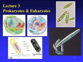
Lecture 3 Structure and Function of Prokaryotes. (Text Chapter: 4.5 - 4.14). Bacterial cell. Cell Membrane. Cell Membrane. Lipid bilayer : hydrophilic exteriors and a hydrophobic interior.

E N D
Lecture 3Structure and Function of Prokaryotes (Text Chapter: 4.5 - 4.14)
Cell Membrane • Lipid bilayer : hydrophilic exteriors and a hydrophobic interior. • Highly selective permeability : The attraction of the nonpolar fatty acid portions of one phospholipid layer for the other layer helps to account for the selective permeability of the cell membrane. • Hopanoids in bacteria (similar to sterols in eukaryotes), may strengthen the membrane as a result of their rigid planar structure. • Integral proteins involved in transport and other functions traverse the membrane.
Prokaryotic vs Eukaryotic Membranes • Different composition of phospholipid • Absence of sterols in prokaryotic membranes • In many bacteria hopanoids instead
Bacteria vs Archaea Membranes • Archaea contain ether-linked lipids • No fatty acids but isoprene (5 carbon hydrocarbon) in Archaea • Some Archaea have lipid monolayers
Monolayer Membrane of Archaea • Monolayer results from covalent linkage of glycerol side chains
Function of Cell Membrane • Permeability barrier • Prevents leakage of cytoplasmic metabolites into the environment • Selective permeability also prevents diffusion of most solutes
Anchor for membrane proteins involved in • Specific transport mechanisms to accumulate nutrients against the concentration gradient • Bioenergetics • Chemotaxis • Site for energy conservation in the cell
The Outer Membrane of Gram-Negative Bacteria • Outer membrane consisting of lipopolysaccharide (LPS), protein, and lipoprotein • Porins allow for permeability across the outer membrane by creating channels that traverse the membrane. • Periplasm, which contains various proteins involved in important cellular functions.
Lipopolysaccharide (LPS) • Lipopolysaccharide (LPS) is composed of lipid A, a core polysaccharide, and an O-specific polysaccharide (Figure 4.34). • Lipid A of LPS has endotoxin properties, which may cause violent symptoms in humans (fever and, if the concentration is high enough, shock).
Cell Wall and Gram Stain • The structural differences between the cell walls of gram-positive and gram-negative Bacteria are thought to be responsible for differences in the Gram stain reaction. • Alcohol can readily penetrate the lipid-rich outer membrane of gram-negative Bacteria and extract the insoluble crystal violet-iodine complex from the thin peptidoglycan layer.
Exceptions: Bacteria without Cell Walls • Some prokaryotes are free-living protoplasts that survive without cell walls because they have unusually tough membranes or live in osmotically protected habitats, such as the animal body. • Species of mycoplasmas have no cell walls because they live in animal cells and they possess stronger membrane due to higher content of sterol, which they assumed from the host.
Cell Walls of Archaea • Pseudopeptidoglycan : Consist of N-acetyl talosaminuronic acid (NAT) • b-1,3 bonds instead of the b-1,4 bonds of peptidoglycan (Lysozyme insensitive)
Bacterial Shape: Common Shapes • Coccus • Round, spherical • E.g. Staphylococcus epidermidis • Rod (bacillus) • E.g. E. coli, B. anthracis • Spiral • Fixed: spirilla • Flexible: spirochetes (Treponema pallidum) • Filamentous
Bacterial Shape: Additional Shapes • Unusual • Star-shaped • Square • Triangular • Pleomorphic • Within a population various shapes (Corynebacteria) • No fixed shape • Cellwall-less: Mycoplasma/Ureaplasma Shape is influenced by environmental conditions, age of culture, antibiotic pretreatment!
Surface Structures • Prokaryotic cells contain various surface structures • Fimbriae (F) and pili (P) • S-layers (S) • Capsules (C) • Slime layers (SL) • Key functions • Attaching cells to a solid surface or other cells (F, P,SL) • Providing protection (S, C)
Surface Structures: Fimbria and Pili • Protein filaments • Shorter and thinner: fimbriae • Longer and fewer: pili • filaments that are best known for their function in conjugation (bacterial sex)
Surface Structures: S-Layer • Two-dimensional array of protein called an S-layer • Often hexagonal symmetry • Selective sieve allowing the passage of low-molecular-weight substances while excluding large molecules and structures • Protection • Mainly in Archaea, but also found in some Bacteria
Surface Structures: Capsules and Slime Layer • Carbohydrates • Protection against desiccation • Capsule • more rigid and thicker • excludes more particles • sometimes made of protein • more protective, antiphagocytic • Slime layer • Attachment
Inclusion Bodies • Prokaryotic cells often contain internal granules that function as energy reserve, reservoir for building blocks, or in magnetotaxis • Lipids, sugars • Poly--hydroxyalkanoates (PHAs) such as poly--hydroxybutyrate (PHB) • Glycogen • Inorganic substances • Polyphosphate • Elemental sulfur (in periplasm) • Magnetite
Inclusion Bodies: Magnetosomes • Intracellular particles of the iron mineral magnetite (Fe3O4) that allow organisms to respond to a magnetic field • Orientation and alignment along Earth’s magnetic field, probably to deeper sediments
Gas Vesicles • Small gas-filled structures made of protein that confer buoyancy on cells by decreasing their density • Contain two different proteins arranged to form a gas-permeable structure impermeable for water and solutes (Figure 4.46) • Means of motility, which allows organisms in water to position themselves for optimum light harvesting • Common in many species of cyanobacteria
cross section longitudinal Gas Vesicles in Cyanobacteria
Endospores • Highly resistant differentiated bacterial cell • Produced by certain gram-positive (Bacillus sp, Clostridium sp) • Enable the organism to endure extreme environmental conditions • Endospore formation leads to a highly dehydrated structure • Contain essential macromolecules and a variety of substances absent from vegetative cells
Endospores (II) • Significantly different from the vegetative, or normal functioning cells • Calcium–diplicolinic acid complexes • Reduce water availability within the endospore thus helping to dehydrate it • Intercalate in DNA and stabilizing it to prevent heat denaturation • Small acid-soluble proteins • Protect DNA from UV radiation, desiccation, and dry heat • Serve as a carbon and energy source during germination • Can remain dormant indefinitely but germinate quickly when the appropriate trigger is applied
Endospores (III) • Emergence of the vegetative cell is the result of endospore activation, germination, and subsequent outgrowth
Flagella • Motility in most microorganisms is accomplished by flagella. In prokaryotes, the flagellum is a complex structure made of several proteins, most of which are anchored in the cell wall and cytoplasmic membrane. • The flagellum filament, which is made of a single kind of protein (flagellin), rotates at the expense of the proton motive force, which drives the flagellar motor. • Flagella move the cell by rotation, much like the propeller in a motor boat (Figure 4.56). An appreciable speed of about 60 cell lengths/second can be achieved.
Hook and basal body differ in gram-positive and gram-negative bacteria!