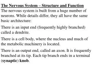
First lecture: Immune system structure and function
First lecture: Immune system structure and function. Immune system Immune system like any other system in the body includes: Organs, tissues, cells, molecules and some times fluids. The tissue of the immune system are called lymphoid tissue.


First lecture: Immune system structure and function
E N D
Presentation Transcript
First lecture: Immune system structure and function
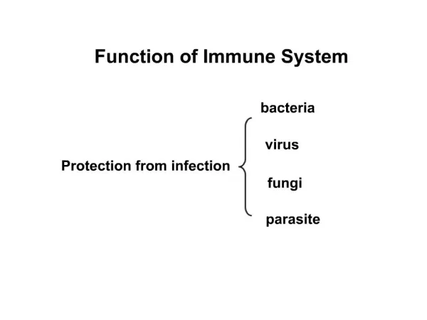
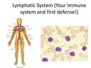
Immune system Immune system like any other system in the body includes: Organs, tissues, cells, molecules and some times fluids. The tissue of the immune system are called lymphoid tissue. The cells of the immune system are all blood cells except the RBCs. The molecules of the immune system are: antibodies. complement, cytokine, chemokines etc. The fluid of immune system is called lymph. The lymph: is plasma components.
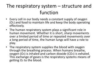
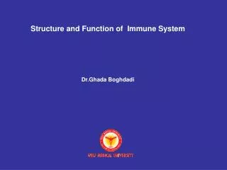
OVERVIEW OF THE IMMUNE SYSTEMOrganized similarly to nervous system: Cells of the immune system (IS) found throughout the body, but also found in specialized organs.Cells:lymphocytes, macrophages & monocytes, dendritic cells,granulocytes. All arise from pluripotent hematopoietic progenitor cells in bone marrow.Organs:lymph nodes (found in various locations), thymus, spleen - these constitute the lymphoid organs.
Immune system organs and tissues (Lymphoid tissue)
(not truly a lymphoid organ, but the source of IS progenitor cells) LOCATION OF MAJOR LYMPHOID ORGANS THROUGHOUT THE BODY
Lymphoid tissues:Are the sites where the ; – Generation, – Maturation, – Habitationand – Activation of the immune cells take place.
Lymphocytes: Major subtypes are T and B cells, responsible for immunological memory. T cells mature in thymus; B cells in avian Bursa of Fabricius but mammalian fetal liver & bone marrow. Cells participate in cell-mediated immunity & regulation responses; B cells synthesize Abs. NK cells are morphologically similar to T & B cells; are cytotoxic in absence of prior stimulation.
lymphocyte from blood smear, Wright-Giemsa stain, 1000x
LOCATION OF THE BURSA OF FABRICIUS T and B cells have specific antigen receptors, which play roles in developing immunological memory and in specificity of the immune response to antigens. Both T and B cells secrete proteins called cytokines, which form the communication system among and between cells and cell types.
T cellsB cells Ag receptor TCR related to Ig BCR is membrane-bound Ig but not Ig Ag recognition in context of MHC can recognize Ag alone on APC or accessory cells Functional Th (helper) and subsets of B cells not subsets Tc (cytolytic) different in function Secrete Cytokines Ig (as Ab) and cytokines Surface CD4 and CD8 Ig (among many others) markers (among many others) When Ag- Become (proliferating) Become lymphoblasts, then activated lymphoblasts become plasma cells Costimulation Yes No required? T versus B Cells
TWO MAJOR TYPES OF T CELLS Th1 & Th2 Effectors cells help B cells make Ab Class I MHC expression – ubiquitous (every where). Class II MHC expression - constitutive: restricted to B cells, a proportion of monos / macs & DCs after activation: induced on most cell types.
mono in blood smear Wright Giemsa, 1000 x macrophage in tissue, H&E stain, 400x MONOCYTES AND MACROPHAGES Monocytes are immature macrophages, circulate in blood & accumulate at sites of inflammation. MQs may differentiate in tissue in absence of antigen (e.g. Kupffer cells in liver) or differentiate in response to Ag. They are Ag-presenting cells (APC) and cooperate with B and T cells in mounting immune responses. Also phagocytose microbes; contain bactericidal mechanisms.
MONOS AND MQS-CONTINUED Express a myeloid receptor (CD14) which serves as a recognition molecule for a wide variety of bacterial envelope molecules, such as LPS from Gram -ve organisms and components of Mycobacterial and Gram +ve cell walls. Ligation of this receptor leads to MQs activation.
Also they’re activated by T cell derived cytokines leading to increased phagocytosis and microbicidal activity (increased activity of degradative enzymes, nitrogen and oxygen free radical production and prostaglandins etc.).
NOTE: T cell derived cytokines increase the antigen presenting activity of macrophages which, in turn, are able to present antigen to T cells. This cycle will continue as a positive feedback loop until the antigen is eliminated.
LPS-activated DCs DENDRITIC CELLS DCs are the APCs for primary (1st time) adaptive immune responses. Also constitute major components of the innate immune system and the bridge to adaptive immunity. Two sites of origin, plasmacytoidDCs (periphery & spleen) and myeloid DCs (bone marrow). So far no major functional differences noted.
THE DENDRITIC CELL THAT’S NOT A DENDRITIC CELL Follicular DCs are found in primary & secondary lymphoid follicles but are not of the same origin as plasmacytoid or myeloid DCs. In fact, their exact origin is unknown, though they may be myeloid. FDCs play a role in controlling B cells responses. FDCs in the spleen
GRANULOCYTESPolymorphonuclear leukocytes (PMNs) or Neutrophils: Predominant type of white blood cell, rapidly migrate to sites of infection or inflammation. Phagocytic, they have special enzymatic pathways for enhanced bactericidal action. Also called azurophils, due to blue-stained (azurophilic) granules.
azurophilic staining of PMNs PMN Mono PMN, note tri-lobed nucleus Wright-Giemsa, 1000x Comparison of mono to PMN
Basophil mast cells Degranulating mast cell intact mast cell BASOPHILS AND MAST CELLHave basophilic granules, which contain mediators, especially of allergic responses. Basophils circulate, mast cells found in tissue.
Eosinophil Eosinophil PMNs Comparison of PMNs to eosinophil EOSINOPHILS Have granules that stain red with eosin. Mediate late phase of allergic response, active in immune response to parasites & tumors (antibody- dependent cell-mediate cytotoxicity). Granules contain toxic proteins of high pH. eosinophil
Hematopoietic stem cell Stromal stem cell ORGANS OF THE IMMUNE SYSTEM PRIMARY LYMPHOID ORGANS Primary lymphoid organs are where lymphocytes arise and mature in the absence of antigenic stimuli. They are the bone marrow and thymus. Bone marrow: Source of all hematopoietic progenitor (stem) cells, site of B cell maturation post-birth in mammals.
Primary lymphoid tissuesThe sites where the blood cells, generation (haematopoiesis) and maturation take place Haematopoiesis takes place:• In yolk sac (first 5 weeks of fetus’s age)• In fetal liver (5-8 weeks of age)• In the whole bone (after 4 months of fetus’s age)In adult the haematopoiesis takes place in the flat bones only (sternum, scapula, skull, pelvis) – Bone marrow: the site where all blood cells generation and maturation (except T cells maturation) take place.
مغزاستخوان : سه فونکسیون به عهده دارد : مرکزتولید اکثررده های سلولی مختلف خونی می باشد . درمغزاستخوان Stem Cell لنفوئید ها را می سازد . ازسلول اخیر سلول های T لمفوئید ، B لمفوئید ، NK Cell یا سلول های کشندۀ طبیعی و K Cell ها مشتق می شوند . سلول میلوئیدی دیگر سلولی است که از Stem cellها مشتق می شود و تمام رده های سلول های خونی ازاین سلول مشتق می شوند ازجمله مگا کاریوسیت ها Megacaryocyte که این ها نیز به نوبۀ خود سلول های خونی نوع پلاکت را می سازند. مونوسیت ها که سازندۀ ماکروفاژها هستند نیزاز Myeloid cellها منشأ می گیرند. دندریتیک سل ها( dendritic cell) که ماکروفاژهای تخصص یافته هستند نیزازاین سلول ها منشأ می گیرند و ماست سل ها mast cells نیز ازاين سلول ها مشتق گردیده اند.
دومین وظیفۀ مغزاستخوان این است که به عنوان مرکزی است برای جذب آنتی ژن به دلیل اینکه دارای مقدارزیادی ماکروفاژمی باشد . مغزاستخوان محلی است برای تولید وذخیرۀ آنتی بادی بطوری که دربعضی موجودات بیش از50% آنتی بادی ها درمغزاستخوان تولید می شوند که بیشتر ازنوع IgG می باشند لذا مغزاستخوان هم می تواند یک ارگان لنفوئیدی اولیه باشد چون همۀ سلول های خونی از آن جا سرچشمه می گیرند وهم یک ارگان ثانویۀ لنفوئیدی زیرا محلی است برای جذب آنتی ژن و پاسخ برعلیه آن . مغزاستخوان دارای دو بخش جداگانه است : الف ) Vascular Compatment ب ) Hematopoetic Compatment بخش H.Com… محل تشکیل سلول های خونی و بخش V.Com… محل عروق خونی ، سینوس های خونی و RBCها ونیزمغزاستخوان است.
PRIMARY LYMPHOID ORGANS: THYMUS The thymus is the site where lymphoid cells undergo maturation and education into T cells prior to release into the circulation. This process allows T cells to develop the important attribute known as self-tolerance. The thymus is found in the thorax in the anterior mediastinum. It gradually enlarges during childhood but after puberty it undergoes a process of involution resulting in a reduction in the functioning mass of the gland. It continues to function throughout life, however. The thymus is arranged into an outer cellular cortex and an inner medulla. Immature lymphoid cells enter the cortex, where they proliferate, mature, and move to the medulla, from where mature T lymphocytes enter the circulation.
هورمون های تیموس : تیموس هورمون های مختلفی تولید می کند وبعنوان یک غدۀ آندوکرین داخلی تلقی می شود ازجمله تیموزینThymosine که دارای سه تیپ آلفا ، بتا و گاما می شود وهرکدام نیزگروهی فرعی دیگری دارند . اثر این هورمون القاء ظهورآنزیم T.D.T (Terminal Deoxynucleotidyl Transferase ) وهمچنین ظهورReceptor گلبول قرمز برسطح لنفوسیت های T که اصطلاحاً به آن Cd2 می گویند می باشند. تیمو پوئی تین Thymopoetin : دو نوع دارد ودرتنظیم پاسخ های ایمنی وافزایش جمعیت لنفوسیت های مهارکننده ( T. Suppressor) دخالت دارد که دربیماری های اتوایمیون Auto Immune ایفای نقش می نماید. Thymic humoral factor : این هورمون می تواند تمایز لنفوسیت های T را تشدید نماید و صلاحیت لنفوسیت های T را درموش های که تیموس آن ها را برداشته اند اعاده نماید. Factor Thymic serige : بوسیلۀ سلول های اپی تلیال تیموس تولید می شود و باعث القاء ظهورآنزیم های سطحی لنفوسیت های T می شود.
تیموس دارای دو بخش است : بخش قشری یا Cortex : ازتعداد زیادی سلول به نام Thymocyte تشکیل یافته است که منشاء آن ها از Pre Thymocyte بوده که این نیز به نوبه خود از Stem Cell درمغزاستخوان منشأ می گیرد. این سلول ها دائماً درحال تقسیم وتمایز بوده وتعدادی از آن ها ازبین می رود وبخشی از آن ها که لازم است تمایزیافته و وارد مدولا Medulla می شود. بخش مرکزی یا Medulla : این بخش ازبافت اپی تلیال تیموس تشکیل یافته است ودراین بخش اجسامی قراردارند که بنام اجسام هاسل یا Hassal body نام دارند . درسلول های اپی تلیال این بخش مقداری لنوسیت دیده می شود . بنظرمیرسد که لنفوسیت ها درحال آموزش هستند وسلول هایی که لنفوسیت درآن ها درحال آموزش هستند بنام سلول پرستاریا CellNurse نامیده می شوند .
اجسام هاسل : بیش از90% لنفوسیت های وارد شده به تیموس ازبین می روند وتشکیل اجسام هاسل را می دهند . درداخل تیموس یک دسته سلول دیگر بنام میلوئید سلول وجود دارد که دارای نقاط تاریک و روشن بوده شبیه سلول های ماهیچه ای می باشند وبنظرمی رسد که Muscular distrophy ( دیستروفی عضلانی ) که درجریان اختلالات تیموس ایجاد می شود به دلیل همین سلول های میلوئیدی است . رگ های خونی که وارد تیموس می شود درناحیۀ کورتکس دارای اندوتلیال و یک لایۀ ، ممبران بازال Membrane basal یا Basment membrane می باشد که این وضعیت همراه وجود ماکروفاژها مانع از ورود آنتی ژن به داخل تیموس می شود . لذا تیموس یک عضو Thymic blood barrier می باشد ، بدین سبب آموزش لنفوسیت ها درداخل تیموس بدون حضورآنتی ژن های خارجی صورت می گیرد.
THYMUS young Cortex - dark Connective tissue Lobules Medulla - light Hassall’s thymic corpuscle Packed lymphocytes (thymocytes) round, red, layered
PRINCIPAL THYMIC CELLS EPITHELIO-RETICULAR CELL NAدVE LYMPHOCYTES (THYMOCYTES) desmosome MF disposing of un-approved thymocyte
Dendritic cells (DC) B cells Myeloid DC (CD11c+, CD11b+, CD8-) CLP T cells Langerhans cells (skin) (CD11c+, CD11b+, CD8+/-, Langerin) Dendritic cells HSC Lymphoid DC (CD11c+, CD11b-, CD8+) Monocytes Plasmacytoid DC (CD11c+, B220+) CMP granulocytes Monocyte-derived DC (CD11c+/-, CD11b+, CD8-) inflammation erythrocytes Megakaryoctes
Ag PRESENTATION IN LYMPH NODES from Itano & Jenkins. Nature Immunology 4, 733 - 739 (2003). Antigen presentation to naive CD4 T cells in the lymph node.
بعضی این ارگان را معادل بورس درپرندگان می دانند که در روی روده ها قرار دارد وشامل دو قسمت است : الف ) درناحیۀ Ieujenum قراردارد ب ) درناحیۀ Illeum و Secum قراردارد. پلاک های پی یر برروی ایلوئوم درهرساعـت 3600000 لنفوسیت تولید می کند که فقط 200000باقیمانده وبقیۀ آن ازبین می روند درحالیکه پلاک های پی یر واقع در ژژنوم درمراحل زندگی جنینی بوجود آمده وتا پایان عمروجود دارند وکارآن ها جذب وپاسخ به آنتی ژن ها است ودرسرراه لنف قراردارند وبصورت ارگان وندول های لنفاوی ثانويه می باشند که مهمترین آن ها طحال است.