Download
1 / 22
260 likes | 463 Views
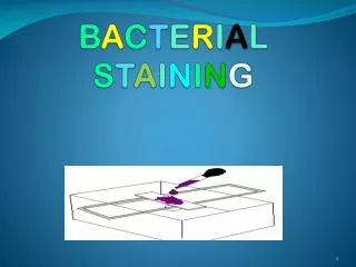
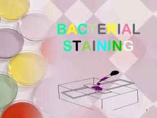
B A C T E R I A L S T A I N I N G. What is a Stain. A stain is a substance that adheres to a cell, giving the cell color. The presence of color gives the cells significant contrast so are much more visible.

E N D
What is a Stain • A stain is a substance that adheres to a cell, giving the cell color. • The presence of color gives the cells significant contrast so are much more visible. • Different stains have different affinities for different organisms, or different parts of organisms • They are used to differentiate different types of organisms or to view specific parts of organisms
Why we should be Stain Bacteria • Bacteria have nearly the same refractive index as water, therefore, when they are observed under a microscope by wet mount they are opaque or nearly invisible to the naked eye. • Different types of staining methods are used to make the cells and their internal structures more visible under the light microscope.
stains composes from • Stains are a mixture of chromogen and auxochrome • Chromogen= benzene derivative + chromophore (coloring agent) • Auxochrome: give +ve or –ve charge to the chromogen • The ionized stain is capable of binding to cell structures with opposite charges.
Basic stains are cationic; when ionized, the chromogeneexhibits a positive charge. Basic stains bind to negatively charged cell structures like nucleic acids. Methylene blue, crystal violet and carbolfuchsinare common basic stains. • Acidic stains are anionic; when ionized, the chromogen exhibits a negative charge. Acidic stains bind to positively charged cell structures like proteins. Picric acid, eosin and nigrosinare common acidic stains. • Bacteria are slightly negatively charged at pH 7.0 • Basic dye stains bacteria • Acidic dye stains background
There are three type of staining in Microbiological lab. • 1. Simple stain. • 2. Differential Stain: (Gram stain , Acid fast Stain) • 3. Special stain: (Capsular stain, Endospore stain, Flagellar stain).
Simple stain • Used to show the general structures (shape and arrangement) of some bacteria. • Aqueous or alcohol solution of single (usually basic) stain is used in this procedure
procedure 1. Obtain broth cultures of the bacteria. • Using an inoculating loop, remove a loopful of suspension from one of the tubes. Remember to use sterile technique. 3. Smear the bacteria across the center of the slide with the loop. If the bacterial suspension is very thick, add a drop of water and mix the bacteria and the water on the slide.
4. Allow the smear to completely air dry. • Air dry first to prevent lysis (boiling) 5. Heat-fix the smear by quickly passing the slide through a Bunsen burner flame three times. This causes partial melting of the cell walls and membranes of the bacteria, and makes them stick to the slide. Do not overheat the slide as this will destroy the bacteria. Heat Fixing • Kill. • Stops autolysis. • Adherence to slide. • Increase dye taking
6. Cover the smear with a few drops of one of the stains. Allow the stain to remain for the following periods of time: Carbolfuchsin- 15-30 seconds. Methylene blue- 1-2 minutes. Nigrosin- 20-60 seconds. • . The stain can be poured drop by drop on the slide 7. Gently rinse the slide by holding its surface parallel to a gently flowing stream of water. 8. Gently blot the excess water from the slide with bibulous paper. Do not wipe the slide. Allow the slide to air dry. Observe the slide under the microscope with air and oil lenses.
Bacterial shape arrangement • Cocci • Bacilli • Clusters (group). • Chains. • Pairs (diploids). • No special arrangement.
Gram stain • Differential stain (Hans Christian Gram, a Danish doctor ). He developed a new method to stain bacteria so they can be visible in specimen samples. • Differentiate bacteria into two large groups (the Gram Positive and the Gram negative) • Gram status is important in medicine; the presence or absence of a cell wall will change the bacterium's susceptibility to some antibiotics • Almost all bacteria are described by their Gram stain characteristics. • Based on differences of Cell wall structures
Reagents for Gram Stain • Crystal Violet (purple). • Primary stain; positive stain • Stains cell wall purple • Iodine • Mordant • Combines with primary stain to form an insoluble complex that gets trapped in bacterial cell wall • Ethanol • Decolorizer • CV-I complex washed out of Gram negative organisms because it cannot be trapped by lippopolysaccharide layer; flows right through outer membrane • Safranin(pink) • Counterstain • Simple positive stain that provides contrasting dye for decolorized cells (Gram negative) • Stains all cells, but only the negative ones actually appear pink.
Gram Staining Procedure (after air drying and heat fixation)
Gm-ve bacilli Gm+vecocci
Errors during staining • Never ever used old culture. • Never ever used sample for patient take antibiotic. • Time of Decolorizer: • Over: G + see as G -. • Low: G- see as G +. • Time of fixation: • Over: G + see as G -. • Low: no sample on slide.