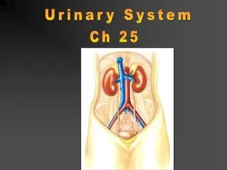
Urinary System
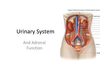
Urinary System. Topics & Objectives. Kidney Anatomy Function Glomerular filtration Tubular reabsorption & secretion Urine excretion & plasma clearance. Kidney Functions:. Ultimately regulate ECF volume (receive ~ 20% of cardiac output!) Maintain H 2 O balance in the body

Urinary System
E N D
Presentation Transcript
Topics & Objectives • Kidney • Anatomy • Function • Glomerular filtration • Tubular reabsorption & secretion • Urine excretion & plasma clearance
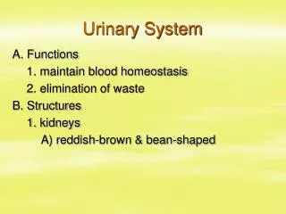
Kidney Functions: • Ultimately regulate ECF volume (receive ~ 20% of cardiac output!) • Maintain H2O balance in the body • Maintain osmolarity • Regulation of ECF ions • Na+, Cl-, K+, H+, etc. • Maintain plasma volume & acid-base balance • Excretion of end products and foreign compounds • Producing EPO & renin
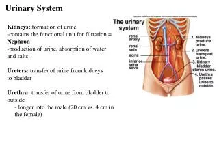
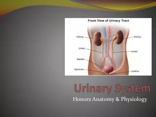
Renal cortex Renal medulla Renal pyramid Renal pelvis Renal artery Ureter Renal vein Kidney Inferior vena cava Aorta Urinary bladder Ureter Urethra Figure 14.1Page 513
Medulla Cortex Nephrons • ~ 1 million within kidney • Functional unit for urine formation • Arrangement comprises renal cortex & renal medulla • Each nephron composed of: • Vascular component • Tubular component
Distal tubule Proximal tubule Collecting duct Juxtaglomerular apparatus Efferent arteriole Afferent arteriole Bowman’s capsule Glomerulus Peritubular capillaries Loop of Henle To renal pelvis Cortex Medulla Figure 14.3Page 514
Efferent arteriole Afferent arteriole Glomerulus Peritubular capillaries Vascular Component: • Glomerulus • Location for H2O and solute filtration from blood • Arterioles • Afferent: to glomerular capillaries • Efferent: drains capillaries • No O2 extraction! • Peritubular capillaries • Supply renal tissue
Distal tubule Proximal tubule Juxtaglomerular apparatus Bowman’s capsule Loop of Henle Tubular Component: • Bowman’s capsule • Collects fluid from glomerulus • Fluid travels to: • Proximal tubule • Loop of Henle • Passes through juxtaglomerular apparatus • Vascular/tubular component • Distal tubule
Cortical nephron Juxtamedullary nephron
Urine Formation • Glomerular filtration (protein-free) • ~ 20% of the plasma (1st step of urine formation) • ~ 50 gallons each day (PV ~ 65x/day) • Tubular reabsorption • Of the 50 gallons filtered, about 98% reabsorbed • Tubular secretion • ~ 80% of the plasma into the peritubular capillaries
Efferent arteriole Afferent arteriole Glomerulus GF Bowman’s capsule TR Peritubular capillary TS Kidney tubule (entire length, uncoiled) 80% of the plasma that enters the glomerulus is not filtered and leaves through the efferent arteriole. 20% of the plasma that enters the glomerulus is filtered. To venous system (conserved for the body) Urine excretion (eliminated from the body) Figure 14.6Page 516
Figure 14.7Page 517 Blood pathway Venous blood Glomerular capillaries Efferent arteriole Peritubular capillaries Glomerular filtration Tubular reabsorption Tubular secretion Filtrate pathway Bowman’s capsule Tubule (from proximal tubule to collecting duct) Urine
Afferent arteriole Efferent arteriole Glomerulus Glomerular capillary Bowman’s capsule Basement membrane Proximal convoluted tubule Figure 14.8 (1)Page 518
Basement membrane Glomerular Filtration Lumen of glomerular capillary Endothelial cell pores podocytes Figure 14.8 (3)Page 518 Lumen of Bowman’s capsule
Glomerular Filtration (cont.): • Occurs through pressure gradients… • Capillary blood pressure (~55mmHg) • Favors filtration • Plasma osmotic pressure (~30mmHg) • Caused by distribution of plasma proteins across glomerular membrane • Cannot cross into Bowman’s capsule • Bowman’s capsule hydrostatic pressure (~15mmHG) • Pressure by the fluid • All three pressures determine filtration rate!
1) Changes in BP 2) Osmotic pressure 3) Hydrostatic pressure Look at Table 14.1!
GFR Regulation • Autoregulation • Prevents spontaneous changes in GFR • Vasoconstriction & vasodilation • Myogenic mechanism – response to stretch • Tubuloglomerular feedback mechanism • Extrinsic sympathetic control • Long-term regulation of arterial BP • Sympathetic nervous system (no parasympathetic activity) • Baroreceptor reflex
Glomerulus Glomerular capillary blood pressure Afferent arteriole Efferent arteriole Arterial blood pressure (increases blood flow into the glomerulus) Net filtration pressure GFR GFR autoregulation • Alterations in arteriolar afferent & efferent blood pressures Figure 14.10Page 520
Glomerular capillary blood pressure Net filtration pressure GFR GFR autoregulation (cont.) Glomerulus Afferent arteriole Efferent arteriole Vasoconstriction (decreases blood flow into the glomerulus)
Glomerular capillary blood pressure Net filtration pressure GFR GFR autoregulation Glomerulus Afferent arteriole Efferent arteriole Vasodilation (increases blood flow into the glomerulus)
Distal tubule Bowman’s capsule Efferent arteriole Afferent arteriole GFR Autoregulation – Tubuloglomerular feedback • Smooth muscle cells within afferent arteriole • Granular cells – secretory capabilities • Tubular cells (macula densa) • Detect changes in the rate of fluid passing through tubule • Bring about vasoconstriction or vasodilation
Efferent arteriole Endothelial cell Lumen of Bowman’s capsule Smooth muscle cell Glomerular capillaries Macula densa Podocyte Granular cells Distal tubule Afferent arteriole
Arterial blood pressure Driving pressure into glomerulus Glomerular capillary pressure GFR Rate of fluid flow through tubules Stimulation of macula densa cells to release vasoactive chemicals Chemicals released that induce afferent arteriolar vasoconstriction Blood flow into glomerulus Glomerular capillary pressure to normal GFR to normal
Extrinsic Control – Baroreceptors • Response to decreased BP • Sympathetically induced vasoconstriction • Afferent arterioles (sympathetically innervated) • Response to increased BP • Sympathetic stimulation decreases
Long-term adjustment for Short-term adjustment for Arterial blood pressure Detection by aortic arch and carotid sinus baroreceptors Arterial blood pressure Cardiac output Sympathetic activity Total peripheral resistance Generalized arteriolar vasoconstriction Afferent arteriolar vasoconstriction Glomerular capillary blood pressure GFR Urine volume Conservation of fluid and salt Arterial blood pressure
Efferent arteriole Afferent arteriole Glomerulus GF Bowman’s capsule TR Peritubular capillary Figure 14.6Page 516
Tubular reabsorption: • Ultimately attempting to maintain body’s internal environment • Proper composition & volume Table 14.2
Peritubular capillary Tubular lumen Tubular epithelial cell Plasma Tight junction 1) Luminal membrane 3) Basolateral membrane 5) Capillary wall • Material must pass through the cells (5 steps) 4) Interstitial fluid 2) Cytosol Figure 14.17Page 526 Transepithelial transport
Constant percentage of Na+ reabsorption Passive & Active Reabsorption • Passive: all steps follow electrochemical or osmotic gradients • Active: any one of the steps requiring energy • Sodium (80% of kidney’s total energy requirement) • ~ 67% in proximal tubule • ~ 25% in loop of Henle • ~ 8% in distal and collecting tubules • Glucose • Phosphate
Peritubular capillary Lumen Tubular cell Interstitial fluid Diffusion Na+ channel Active transport Basolateral Na+– K+ ATPase carrier • Na+ pumped out against concentration gradient • Creates higher concentration in interstitial fluid & allows for passive diffusion back into lumen Diffusion Figure 14.18Page 527
Distal tubule • Distal tubule (~ 8% of total reabsorption) is hormonally regulated • Related to total Na+ load in body • Changes in ECF affect osmotic pressures ex: Increased Na+ in ECF causes increased H2O in ECF • Ultimately regulates blood pressure • Renin-angiotensin-aldosterone system ↑ • Atrial natriuretic peptide ↓
Within juxtaglomerular apparatus… • Granular cells release renin • In response to fall of NaCl/ ECF volume/ BP • Recognized by intrarenal baroreceptors • Sympathetic response to secrete more renin • Ultimately trying to increase plasma volume
Figure 14.19Page 529 NaCl / ECF volume / Arterial blood pressure Na+ reabsorption by kidney tubules ( CI– reabsorption follows passively) H2O conserved Adrenal cortex Liver Kidney Lungs Kidney Na+ (and CI–) osmotically hold more H2O in ECF Na+ (and CI–) conserved Angiotensin- converting enzyme Renin Angiotensinogen Angiotensin I Angiotensin II Aldosterone Vasopressin Thirst Arteriolar vasoconstriction H2O reabsorption by kidney tubules Fluid intake
Helps correct Helps correct Figure 14.20Page 530 NaCl / ECF volume / Arterial blood pressure Cardiac output Total peripheral resistance GFR Arterial blood pressure Na+ excretion in urine Na+ and H2O filtered H2O excretion in urine Cardiac atria Atrial natriuretic peptide Salt-conserving renin-angiotensin- aldosterone system Na+ reabsorption by kidney tubules Sympathetic nervous system Smooth muscle of afferent arterioles Afferent arteriolar vasodilation Inhibits aldosterone & renin secretion
Glucose and amino acid reabsorption • Na+ dependent secondary active transport • Co-transporters that do not require energy • Maximal reabsorption rate depends on substance
Cotransport carrier No energy required Luminal border Energy required Na+–K+ pump No energy required Glucose carrier Basolateral border Blood vessel
Phosphate & Calcium reabsorption • Dependent upon total body content • Regulated by kidneys • Hormonally (parathyroid hormone) • Na+ reabsorption responsible for passive reabsorption of Cl-, H2O, and urea
Peritubular capillary Lumen Proximal tubular cell Interstitial fluid Water channel • H2O (passive) reabsorption • 80% in proximal tubules & loops of Henle Osmosis Figure 14.22Page 533 Hydrostatic pressure Osmosis
Glomerulus Peritubular capillary Bowman’s capsule Beginning of proximal tubule End of proximal tubule = Urea molecules • Urea (passive) reabsorption • Waste product of protein • Becomes increasingly concentrated 125 ml of filtrate Na+ (active) H2O (osmosis) Na+ (active) H2O (osmosis) 44 ml of filtrate Figure 14.23Page 534 Passive diffusion of urea down its concentration gradient
Figure 14.6Page 516 Efferent arteriole Afferent arteriole Glomerulus GF Bowman’s capsule Peritubular capillary TS Kidney tubule (entire length, uncoiled)
Tubular secretion: • Hydrogen ions (H+) • Acid-base regulation throughout the body • Potassium (K+) • Early reabsorption into tubules not regulated • Secretion in distal tubules regulated • Na+-K+ pump • Aldosterone • Organic anions & cations • Foreign compounds, chemical messengers
Figure 14.25Page 536 Na+/ ECF volume/ arterial pressure Renin Angiotensin I Plasma K+ Angiotensin II Aldosterone Tubular K+ secretion Tubular Na+ reabsorption Urinary K+ excretion Urinary Na+ excretion Aldosterone - dual regulation 1) Na+ & K+
Fig. 14.26a Page 539 • Substance: • Filtered • NOT reabsorbed • NOT secreted Peritubular capillary Glomerulus All filtered plasma is cleared of substance Tubule In urine
Substance: • Filtered • NOT secreted • Completely reabsorbed None of filtered plasma is cleared of substance
Substance: • Filtered • NOT secreted • Partially reabsorbed Portion of filtered plasma is cleared of substance