Download
1 / 29
290 likes | 431 Views
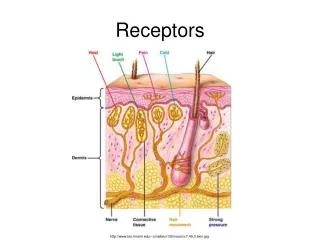
I. Receptors A. All sensory receptors are transducers of some sort, that is, they change an incoming stimulus of pressure, vibration, light, etc., into electro-chemical neuron impulses. Each is specific in that it can transduce only certain types of stimuli into neuron action potentials.

E N D
I. Receptors • A. All sensory receptors are transducers of some sort, that is, they change an incoming stimulus of pressure, vibration, light, etc., into electro-chemical neuron impulses. Each is specific in that it can transduce only certain types of stimuli into neuron action potentials. • B. Perception - conscious awareness of a sensation • C. Interpretation of Sensory Information • 1. _receptive field________ - that region monitored by a receptor cell
D. Central Processing and Adaptation • 1. _adaptation__ - loss of sensitivity after exposure to a stimulus. - • 2. _sensory adaptation_ - result of sensor fatigue , receptor becomes less sensitive to stimulus - smell has great adaptation E. Sensory Limitations • 1. there are many stimuli for which we do not have receptors - • 2. stimuli may be out of range for the appropriate receptors - uv light for example - KESTREL, • Dog whistle • 3. our awareness depends on interpretation which may be inexact.
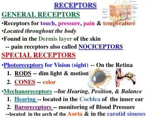
II. General Senses • A._Exteroceptors_- detect the external environment (temperature, pressure, light, etc.) • B. _Interoceptors - provide information about internal conditions, most often tension or stretch in smooth muscle or • tendons. • 1. nociceptor stimulation results in pain. heat, mechanical stress and chemicals can allc ause pain by stimulating different classes of receptors. • a. _fast pain- deep cuts, etc. - referred immediately to primary sensory cortex, usually trigger reflexes • b. __slow pain_ - later, persistent, indistinct source, thalamic • c. _referred pain - visceral, "incorrect" source perceived • 2. _thermoreceptors_ - detect heat or absence thereof, phasic, same pathways as pain • 3. _mechanoreceptors - stimulated by anything moving or physical perturbation. • a. __tactile receptors _ - touch, pressure, vibration b. _baroreceptorsare sensitive to internal pressures blood pressure, lung stretch, digestive tract tension c. __proprioceptors_ are kinesthetic monitors of tendon muscle stretch. • 4. _chemoreceptors- respond to specific chemicals either in solution (taste) or volatilized (smell) or internally in monitoring blood composition. Internal chemoreceptors monitor blood composition - Na+, pH, osmolarity, etc.
III. Special senses • A. _Olfactory (Smell) • distant chemical sense, many chemicals can be detected at a distance as long as they are gaseous. • 1. receptors -sensory epithelium, olfactory receptors and glands, supporting cells and basal cells - covers superior nasal conchae and septum.
B. _Gustation_ (Taste) - taste is chemical also but requires that the dissolved chemicals make contact with the tongue or mouth. • 1. gustatory receptors - gustatory __chemoreceptors_____on the sides of taste buds. Taste buds are incorporated in the papillae which are described by their shapes, • a. foliate - thin, thread like projections • b. _fungiform- shaped like mushrooms. • c. _circumvallate- large target-shaped bumps near the back of the tongue.
2. gustatory pathways - cranial nerves VII, IX and X to the nucleus solitarius in medulla oblongata to gustatory cortex • 3. gustatory discrimination - six tastes • a. sour - (H+) • b. sweet - (organic) • c. salt - (metallic) • d. bitter - (alkaloids) • e. water • f. umami - (savory)
C. Equilibrium and Hearing – ears - MECHANORECEPTORS • 1. external ear • a. pinna – (auricle) funnels sound waves down the external auditory canal (where ceremen “ear wax” is found – helps lubricate and protect opening from bacteria, fungi etc..) • b. tympanic membrane – vibrates and transmits sound to malleus (bone) of middle ear
2. middle ear - three bones and space that amplify and transmit sound to the oval window of inner ear. a. auditory (Eustachian) tube - connects throat and middle ear – (ear/throat infections) • b. auditory ossicles - amplify force of vibration • i. malleus (hammer) – 1st bone • ii. incus (anvil) – 2nd bone • iii. stapes (stirrup) – 3rd bone • c. tensor tympani muscle - stiffens tympanic membrane • d. stapedius muscle - dampens movement of stapes
3. inner ear • a. labyrinth - membranous and bony - is a series of bones and membranes (line bones) which consists of the cochlea (hearing) and vestibule (balence( • i. endolymph & perilymph - fluid that is found inside the triggers hair cells to move, and therefore send nerve impulse
b. cochlea • i. cochlear duct long tube that is rolled like snail shell containing the • ii. Organ of Corti – group of cells devoted to respond to vibration • iii. tympanic & vestibular ducts, fluid filled ducts on either side of organ of corti that carries sound • iv. tectorial membrane, - membrane that receives vibration and stimulates the hair cells • v. hair cells with stereocilia - cells that trigger impulse when touched • vi. round window - allows vibrations to leave and occur • vii. oval window - begins vibrations from stapes
c. semicircular canals • i. three mutually perpendicular- posterior, lateral and anterior • ii. ampulla - where ends of canals meet vestibule • iii. maculae - hair cells with otoliths – cupula is gel –like fluid on hair cells. As head moves if moves the cupula, which bends the hair a certain direction – triggering movement sensation
IV.. Vision – eye - photoreceptors • A. Accessory Structures • 1. eyelids – palpebrae, some of the thinist skin in human body a. medial ( with lacrimal caruncle) and lateral canthus - corners of eyes • b. eyelashes - with sebaceous glands (of Zeis) • c. conjunctiva - mucous membrane lining eyelids • 2. lacrimal apparatus • a. _ lacrimal gland_ - superior and lateral to eye • b. lacrimal puncta - holes near nose to drain tears • c. _lacrimal canaliculi- drain tears to • d. nasolacrimal ducts_ - empties to nasal cavity a = lacrimal glandb = superior lacrimal punctumc = superior lacrimal canald = lacrimal sace = inferior lacrimal punctumf = inferior lacrimal canalg = nasolacrimal canal
B. Eye • 1. superficial structures 3 sets of antagonistic muscles that move eye • a. cushioned by fat pads • 2. fibrous tunic - tough outer layer Outer Layer • a. sclera - white part of fibrous tunic • b. cornea - transparent avascular anterior part - window of the eye, protection and adds curvature to focus light - • 3 vascular tunic -(uvea) • a. choroid - heavily vascular • b. iris with pupil hole, circular sphincter muscles and radial dilator muscles • c. _ciliary body - muscles attached to suspensory ligament, regulates focus of lens
4. nervous tunic • a. retina - innermost layer of eye, has photoreceptors • 1. outer pigmented layer -cyanolabe, erythrolabe, chloroplabe, rhodopsin • 2. inner layer -photoreceptors • a) _rods - black/white vision, motion detection • b) _cones_ – color vision, intense light • b. ora serrata - anterior edge of retina c. __macula lutea_ – fovea centralis - all cones, best vision • d. _optic disc_ – blind spot, where optic nerve exits eye e. optic nerve - Cranial Nerve II – takes impulse to occipital cerebral cortex
5. lens • a. ciliary body - composed of ciliary muscle and ciliary ligament, when it relaxes the lens flattens and you see distance better b. suspensory ligament - hold lens in place • c. anterior chamber with _ aqueous humor - watery substance that baths the cornea • d. posterior chamber with _vitreous body (humor) - “snot like material” that helps give the eye its shape
V. Visual Pathways • ___Optic Chiasma_ - optic nerves partially cross in sphenoid • (right side of the field of each eye combining and going to the • lateral geniculate (knee) on the right, those from the left to • the left, of the thalamus which then relays the data to the • visual centers. ) • VI. Cortical Integration • The visual cortex must combine and integrate the incoming • information into a composite which is perceived as depth. • (This is a remarkable feat when one considers that the image • is tiny, curved, inverted, the two images slightly out of • frame, and each missing a large portion at opposite edges • of the field and having a hole in the image.)