Download
1 / 1
10 likes | 181 Views
Successful Treatment of Cervical Ectopic Pregnancy with a Multi-dose Methotrexate Regimen Kiarra King, MD, Carlos Fernandez, MD and Stephen Locher, MD Department of Obstetrics and Gynecology, Advocate Illinois Masonic Medical Center. Case Report. References. Abstract. Conclusion.

E N D
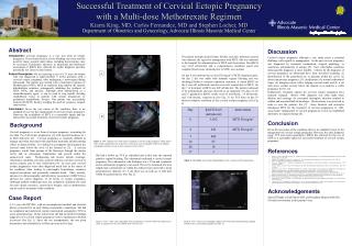
Successful Treatment of Cervical Ectopic Pregnancy with a Multi-dose Methotrexate Regimen Kiarra King, MD, Carlos Fernandez, MD and Stephen Locher, MD Department of Obstetrics and Gynecology, Advocate Illinois Masonic Medical Center Case Report References Abstract Conclusion Discussion Introduction: Cervical pregnancy is a very rare form of ectopic pregnancy. If not detected early, severe bleeding can occur and the need for major surgical intervention, including hysterectomy, may be necessary. Fortunately, advances in ultrasound and laboratory assessment of βHCG have allowed for earlier diagnosis and thus potentially less invasive intervention. Patient Description:We are reporting a case of a 33 year old woman who was diagnosed at approximately 5 weeks gestation with a cervical ectopic pregnancy after undergoing a transvaginal dating ultrasound. The patient was treated with a multi-dose regimen of Methotrexate (MTX). MTX an antifolate drug inhibits the enzyme dihydrofolate reductase subsequently inhibiting the synthesis of DNA, RNA, and proteins. Although often administered as a chemotherapeutic agent, it can be used to stop rapidly dividing trophoblastic tissue in patients with ectopic pregnancies or gestational trophoblastic disease. Our patient was successfully treated with MTX, thereby avoiding the need for extensive surgical intervention. Conclusion: Given the rare nature of the condition, there is no standard of care for the management of cervical ectopic pregnancy. However, the availability of MTX is a reasonable option and has allowed for successful treatment of cervical ectopic pregnancy. The patient strongly desired future fertility, and after informed consent was obtained, she agreed to management with MTX. She was admitted to the hospital for administration of MTX and observation. Her βHCG was 3,432 mUnits/mL and a comprehensive metabolic panel and complete blood count, obtained prior to MTX, were normal. On day 0 of treatment she received 50 mg/m² of MTX intramuscularly. On day 2 she was stable with minimal vaginal bleeding and was discharged home to continue outpatient treatment. A repeat βHCG on day 4 was 643 mUnits/mL and she received a second dose of MTX. On day 7 of treatment a βHCG was 409 mUnits/mL. The patient continued to be asymptomatic and was observed as an outpatient. On days 14, 20 and 27 quantitative βHCG results were 65 mUnits/mL, 14 mUnits/mL and 3 mUnits/mL, respectively (See Fig. 3). An TVS done on day 39 showed complete resolution of the cervical ectopic pregnancy (See Fig 4). EO Cervical ectopic pregnancy although a rare entity poses a potential challenge with regards to management. In the past cervical pregnancy was diagnosed by bimanual examination, surgical pathology, or sometimes, unfortunately, at autopsy [5]. Now, with higher resolution ultrasound the diagnosis is more feasible. Criteria for the diagnosis of cervical pregnancy on ultrasound have been described including (1) identification of the gestational sac or placenta within the cervix (2) absent intrauterine pregnancy; (3) visualization of a normal endometrial stripe; (4) hourglass uterus with a bulging cervical canal, and (5) a sac with active cardiac activity below the internal os to indicate a viable pregnancy [6,9,11,12]. Furthermore, treatment options for cervical ectopic pregnancy have radically changed. Before 1980, diagnosis was often made when dilation and curettage for presumed incomplete abortion resulted in sudden and uncontrollable hemorrhage. Hysterectomy was practiced in order to save the patient's life [7]. Since Farabow and associates introduced MTX for the treatment of cervical pregnancies in 1983, conservative management of cervical pregnancies is now an established alternative to surgical therapy [8]. IO A B Background Given the rare nature of the condition, there is no standard of care for the management of cervical ectopic pregnancy. However, the early diagnosis using TVS and serum quantitative βHCG has allowed for the use of MTX as a reasonable option for successful treatment of cervical ectopic pregnancy. Cervical pregnancy is a rare form of ectopic pregnancy, accounting for less than 1% of all ectopic pregnancies [1]; with reported incidence of 1 in 8628 deliveries [2]. Cervical pregnancy is extremely difficult to manage and often associated with significant morbidity and devastating effects on future fertility. It is defined as a pregnancy developing in the cervical canal below the level of the internal os [3]. A cervical pregnancy results from passage of the blastocyst through the uterine cavity and its subsequent implantation and growth within the endocervical canal. Predisposing risk factors include curettage, Asherman's syndrome, previous cesarean delivery, previous cervical or uterine surgery, and in vitro fertilization [4]. In years past cervical ectopic pregnancies were often diagnosed much later in the course of the condition, often leading to catastrophic hemorrhage, extensive surgical procedures and potentially maternal death. More recently, advances in ultrasonography and laboratory assessment of βHCG have allowed for earlier diagnosis of all forms of ectopic pregnancy. Although modern technology does not completely eliminate the need for more drastic measures, conservative therapy, such as methotrexate can be used for treatment of this condition. Figure 1. TVS - Sagittal view of the uterus visualizing a cervical ectopic pregnancy (white arrow) and a normal endometrial echo (red arrow) with A) gray scale sonography and B) color Doppler sonography. Internal cervical os (IO), external cervical os (EO). She had a follow up TVS as scheduled and at this time she reported painless vaginal bleeding. The ultrasound confirmed a cervical ectopic pregnancy. The endometrial echo thickness was 1.37cm and symmetric and no intrauterine pregnancy was noted. The cervix measured 4.8cm in length, had a gestational sac within the endocervical canal with a mean gestational sac diameter of 1.5 cm; there was no yolk sac or fetal pole within the gestational sac (See Fig. 2). Bouyer J., Coste J., Fernandez H., Pouly J.L., Job-Spira N. Sites of ectopic pregnancy: a 10 year population-based study of 1800 cases. Hum Reprod 2002; 17:3224–3230. Ushakov F.B., Elchalel U., Aceman P.J., Schenker J.G. Cervical pregnancy: past and future. Obstet Gynecol Surv 1997; 52:45–59. Katz V. Comprehensive Gynecology 5th Edition, 2007; 389. Verma U., Maggiorotto F. Conservative management of second-trimester cervical ectopic pregnancy with placenta percreta. Fertil Steril 2006; 87:697. Jeong,E., Kim,Y., Kim H. Triplet Cervical Pregnancy Treated with Intraamniotic Methotrexate. Obstetrics and Gynecology 2002; 100: 1117-1119. Kung F.T., Lin H., Hsu T.Y. Differential diagnosis of suspected cervical pregnancy and conservative treatment with the combination of laparoscopy-assisted uterine artery ligation and hysteroscopic endocervical resection. Fertil Steril 2004; 81:1642-49. Starita A., Di Miscia A., Evangelista S. Cervical ectopic pregnancy: clinical review. Clin Exp Obstet Gynecol 2006; 33:47-49. Farabow W.S., Fulton J.W., Fletcher V., Velat C.A., White J.T. Cervical pregnancy treated with methotrexate. NC Med J 1983; 44:91–93. Bhatt S., Ghazale H., Dogra V. Sonographic Evaluation of Ectopic Pregnancy. Radiologic Clinics of North America 2007; 45:549-560. Mayberger, H. Cervical Pregnancy: A Case Report. Obstetrics and Gynecology 1958; 11: 657-660. Hofmann H.M., Urdl W., Hofler H. Cervical pregnancy: case reports and current concepts in diagnosis and treatment. Arch Gynecol Obstet 1987; 241: 63-69. Timor-Tritsch I.E., Monteagudo A., Mandeville E.O., et al. Successful management of viable cervical pregnancy by local injection of methotrexate guided by transvaginal ultrasonography. Am J Obstet Gynecol 1994; 170:737-394. Figure 3. Chronologic assessment of quantitative β-hCG in relation to MTX administration. CERVIX Acknowledgements FUNDUS Special Thanks to Jude Duval M.D. and Josephine Bunyon R.D.M.S. For their involvement in this patient’s care. A 33 year old G2P1001, with an uncomplicated medical and obstetric history, presented for an early dating sonographic examination. She had no complaints and underwent routine transvaginal sonography (TVS) to assess gestational age. On her initial exam she had inconclusive findings suggestive of a cervical ectopic pregnancy versus a spontaneous abortion in process (See Fig. 1). Since she was asymptomatic, she was given precautions and scheduled for a follow up exam in five days. B A B B Figure 2. TVS – Gray scale sonography of the gestational sac (arrow) within the endocervical canal. A) sagittal view of cervix and B) axial view of cervix. Figure 4. TVS – Gray scale sonography, sagittal view of the uterus demonstrating complete resolution of cervical ectopic pregnancy on day 39.