Ectopic Pregnancy
Ectopic Pregnancy. A’asem Zeidan Abu- Shtaya. Normally, . Normally, . Fertilization occurs in the lateral third of the fallopian tube. On average it takes the spermatozoa 1 hr to reach the ovum.


Ectopic Pregnancy
E N D
Presentation Transcript
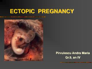
Ectopic Pregnancy A’asemZeidan Abu-Shtaya
Normally, • Fertilization occurs in the lateral third of the fallopian tube. • On average it takes the spermatozoa 1 hr to reach the ovum. • 5-6 days post fertilization, the fertilized egg travels back to the uterus for IMPLANTATION to occur INSIDE the uterine cavity.
Definition • An ectopic pregnancy is one that implants outside the uterine cavity (not the uterus) in a place other than the endometrium
Then.. • Implantation occurs in the fallopian tube in up to 98% of cases • 2nd comes the abdominal implantation, where the placenta could attach to the bowel. With an incidence of ~ 1% • 3d: ovarian ~0.2% • 4th : cervical ~0.2%
Incidence • The incidence of ectopic pregnancies has increased dramatically during hthe past 10 years and now occurs in more than 1:100 pregnancies. • This is thought to be secondary to the increase of: • STI’s • Assisted fertility • PID’s
Why does it happen? • several risk factors predispose patients to extrauterine implantation. • Many affect the fallopian tubes leading to either: • Tubal scarring • Decreased peristaltic motility of the tube.
However.. • in as many as one thirdto one halfof ectopic pregnancies, no risk factors can be identified.
Through the R.F’s • PID’s can lead to intra or peritubal adhesions decreasing the ability of the fertilized ovum to reach the uterine cavity. • STI’s can directly destroy the cilia of the fallopian tube epithelium. • Any tubal surgery like Tubal reanastamosis, tubal ligation, tubal adhesions can cause impairement of cilial movement. • Methods of contraceptions as IUCD prevent the implantation into the endometrial tissue. • Progesterone only pills cause EP because they lead to relaxation of the muscle layer of the tube.
.. • Any inflammatory process within the uterus or the pelvis in general could theoretically lead to the occurrence of ectopic pregnancy. This results from the build-up of scar tissue in the Fallopian tubes, causing damage to cilia.
CLINICAL MANIFESTATIONS • Clinical manifestations typically appear six to eight weeks after the last normal menstrual period, but can occur later, especially if the pregnancy is not in the fallopian tube. Normal pregnancy discomforts (eg, breast tenderness, frequent urination, nausea) are often present.
Diagnosis • History • Physical examination • Lab tests • U/s
History • The 3 classical symptoms of ectopic pregnancy are: • Amenohrea • Abdominal pain • Vaginal bleeding
History • In any lady presenting with those symptoms during her first trimester, EP should be excluded. • you should also think of: • Normal intrauterine pregnancy • Spontaneous abortion • Salpingitis • appendicitis
Physical Examination • Usually associated with minimal findings but may reveal: • Tender adnexal mass • A uterus that is small for gestational age • Cervical bleeding. Patients with RUPTURED EP may be hypotensive, unresponsive, or show signs of peritoneal irritations sec. to haemoperitoneum.
(β-hCG) • EXTREMELY important • Lab method of a choice for confirming EP In normal intrauterine pregnancy (β-hCG) is secreted by the trophoblastic tissue in a predictable manner. The absolute value doubles approximately every 2.5-3 days.
However • In EP the levels are low for gestational age. • e.g. (β-hCG) of 500 IU/ml at day one repeated 3 days later and revealed a value of 2000 IU/ml decreases the likelihood of EP
Progesterone levels • Good specificity and sensitivity for normal intrauterine pregnancy. • Not reliable • If its low, it’s a marker of abnormal pregnancy • Cannot differentiate b/w EP and abortion
U/S • Might show normal IUP, hence we are most probably dealing with an abortion. • might show an adnexal mass • Fetal heart activity in the adenxia could be monitored • Bleeding in doglus pouch
Diagnostic method of a choice is: LAPROSCOPY
What we’re afraid of ?! • There’s always a small risk for heterotropic pregnancy, a multiple gestation with at least one IUP and one EP. • This is a particular concern in the setting of IVF when more than one embryo was utilized • Those patients are labelled as rule-out ectopic. • Should be followed with B-hCG levels every 48 hours and undergo a transvaginal U/s
Management (1) • Mrs. Amireh, a 28 y/o lady presents to the ER with unilateral lower abdominal pain. Fresh vaginal bleeding of 1 day duration. Her LMP was 10 weeks ago. • The Pt. is concious, oriented and looks well. • Upon P/E her pulse was 85, Bp: 130/ 85 Temp. 36.8 • Urine pregnancy test was performed and was positive. • The rotating dr. decided to perform an abdominal U/S that revealed a mass measuring 3 cm in the right fallopian tube and fluid collection in the pouch of doglus . No IUP
We take into consideration: • 1. Patient stability (immediate surgical interferance if unstable) • 2. desire of future fertility • Site of E/P • State of EP (ruptured or intact)
Our very first priority is to stabilize the patient and look for signs of distress. • Our patient looks fine, stable and oriented. • Hence, can be treated either: • Medically • Surgically
Medical Tx: • The drug of a choice used at most institution is MTX • Its appropriate for who have small E/P less than 4 cm and for those patients who are reliable for follow up. • The drug is given as one shot IM 50gm • Patient should be followed with serial b-hCG. • B-hCG levels will rise the first few days after MTX, but will start decreasing after 4-7 days. • If such a fall is not achieved or if the levels plateau, a 2nd dose should be given. • If it stays high MUST go for surgery
Management (2) • Mrs. Badran, a 25 y/o lady, presented to the ER at 4 a.m with severe diffuse abdominal pain. She reports profuse vaginal bleeding. Her last LMP was 8 weeks ago • Upon P/E her Pulse was 110, Bp. 100/70 temp 37 C. severe abdominal tenderness and guarding upon palpation. • The pt is dioriented, drowsy and almost fell to the ground when entering the ER. • Urine b-hCG was positive, U/s showed a ruptured left fallopian tube and severe bleeding within the peritoneum
Surgical Tx: • If a patient presents with a ruptured ectopic pregnancy and is unstable, the first priority is to stabilize with IV fluids, blood products, and pressors if necessary. • The pt should be taken to the OR where exploratory laprotomy can be done to stop the bleeding and remove the ectopic pregnancy
If the pt is stable, the procedure of choice at most institutions is explotatorylaproscopy that can be performed to evacuate the hemoperitoneum , coagulate any ongoing bleeding and resect the ectopic pregnancy
Resection can be either through: • Salpingostomy: the EP is removed leaving the fallopian tube in place • Salpingectomy: where the entire EP along with the tube are removed. * Ovarian EP are normally treated surgically by oophorectomy
Miscarriage A’asemZeidan Abu-Shtaya
Definition also known as spontaneous abortion. refers to a pregnancy that ends spontaneously before the fetus has reached a viable gestational age. The World Health Organization defines it as expulsion or extraction of an embryo or fetus weighing 500 g or less from its mother. This typically corresponds to a gestational age of 20 to 22 weeks or less
Incidence Most common gyanecological and obstetric disorder About 15-25% of all clinically recognized pregnancies. Underestimated because losses that occur 4 to 6 weeks gestational age are often confused with late menses Real incidence could reach 30-35%
First trimester abortions • 60-80% are due to fetal chromosomal abnormalities. (m.c.c) • Incidence of these abnormalities is increased with increasing maternal age. • This could be because many abortions likely occur before implantation. • Other causes: • Infections : genital tract infections and systemic infections with pyrexia • Maternal anatomic defects: uterine anomalies, submucous fibroid, asherman syndrome etc.. • Endocrine and immunological factors • Multiple pregnancy • Cigarette smoking • Psychological disorders All together comprise the remaining 20-30%
Second trimester abortions • Between 13 to 26 weeks gestational age • Less common than first trimester abortions • Have multiple etiologies: • Genital tract infections and PROM • Maternal uterine or cervical anatomic defects • Maternal systemic diseases • Cervical incompetance • TRAUMA • Multiple pregnancy.
Types • Defined by • 1.any or all of the products of conception have passed • 2. whether the cervix is dilated or not
Types ► Threatened abortion ► Inevitable abortion ► Incomplete abortion ► Complete abortion ► Missed abortion ► Septic abortion ► Recurrent aborion
Threatened abortion Any vaginal bleeding before 20 weeks, without dilatation of the cervix or expulsion of any POC (i.e. a normal pregnancy with bleeding)
Threatened abortion- Management 1. reassurance: if fetal heart activity is present, in more than 90% pregnancy will progress in a satisfactory way 2. advice to decrease physical activity and avoid intercourse 3. Give progesterone and hCG which are used in the first trimester to support pregnancy 4. adequate dose of anti-D should be given to all Rh –ve non-immunised patients, whose husbands are Rh +ve 5. label as high risk patients because those patients are liable to late pregnancy complications such as APH and preterm labour.
Inevitable abortion Inevitable heavy vaginal bleeding and dilatation of the cervix WITHOUT expulsion of any POC
Incomplete abortion Incomplete heavy vagial bleeding and dilatation of the cervix WITH partial expulsion but not all the POC
Inevitable and Incomplete - Management Hb, blood grouping, XM 2 units of blood Resucitation, large IV line, fluids and blood transfusion. Those types of abortions can be allowed to finish on their own, with expectant management Or: Can also be taken to completion by either D&C or adminstration of prostaglandins (misoprostol) to induce cervical dilatation and uterine contractions
Complete abortion Complete expulsion of all POC before 20 weeks gestation
Complete -Management 1. conservative management if the uterine cavity is empty Evacuation and curettage in the presence of RPOC Post abortion management
Missed abortion Death of the embryo or fetus before 20 weeks gestation with complete retention of all POC Mostly diagnosed accidentaly during routine U/S in early pregnancy