Download
1 / 12
120 likes | 408 Views
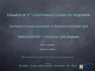
Evaluation of “C” Limbal Relaxing Incisions for Astigmatism Correction in Eyes Submitted to Phacoemulsification and SN6D3 ReSTOR Intraocular Lens Implants. Mario J. Carvalho Giuliano O. Freitas. The authors have no financial interest on this presentation. Setting

E N D
Evaluation of “C” Limbal Relaxing Incisions for AstigmatismCorrection in Eyes Submitted to Phacoemulsification andSN6D3 ReSTOR Intraocular Lens Implants Mario J. Carvalho Giuliano O. Freitas The authors have no financial interest on this presentation Setting ISO Olhos – Ocular Health Institute - Uberlandia – MG - Brazil
Introduction Phacoemulsification’s (PHACO) key aim is fast visual recovery associated to minimal surgical risks (1). Near and distance visual recovery is dependent upon appropriate intraocular lens (IOL) selection (2-5) and intraoperative astigmatism management. Lower surgically induced astigmatism derived from currently available incisions, followed by the implantation of bifocal IOLs have deeply changed cataract surgery focus to a more refractive status, sometimes being referred to as phacorefrative surgery, or also phacorefractive lensectomy (8-9). So, spectacle dependence for near and distance vision has been tremendously lessened by bifocal IOLs, adding an important therapeutic option for cataract surgeons. Fifteen to 20 percent of PHACO patients present topographic astigmatism ranging from 1 to 3 cylinder diopters (CD). Such patients are not good candidates for such an approach, unless preexisting astigmatism is appropriately managed (10). In this scenario, limbal realxing incisions (LRIs) are broadly used to correct corneal topographic astigmatism, due to their easiness to be performed and low cost. LRIs on the other hand, may determine astigmatism hypocorrection, or induce a topographic pattern alteration referred to as adjacent coupling. An alternative to minimize topographic changes sencondary to “conventional” LRIs has been proposed by Carvalho: the “C” limbal relaxing incisions (CLRIs). The present study’s objective is to evaluate CLRIs efficiency, safety and stability in terms of corneal astigmatism reduction among eyes submitted to PHACO followed by bifocal ReSTOR IOL implantation.
Methods The present study has been conducted from September 2004 to July 2008. Patients’ selection has been based on the following criteria: age equals to, or greater than 40 years, cataract occurrence worsening best corrected visual acuity, regular corneal topographic astigmatism ranging from 1 to 2.5 CD and finally, no other ocular or systemic disease that could adversely limit final visual outcomes. Patients’ preoperative evaluation relied on: uncorrected and best corrected visual acuity measurements, anterior segment biomicroscopy at slit lamp, applanation tonometry, indirect binocular ophthalmoscopy, computerized videokeratoscopy (EyeSys 2000 Corneal Analysis System –Eye Sys/ Premier Laser Systems, Inc.; Irvine, CA, U.S.A.,Orbscan, Bausch & Lomb, U.S.A) and immersion biometry (OcuScan XP, Alcon, Forth Worth,TX, U.S.A.). Lens power calculations have took into account simulated keratometry for the central 3 mm optical zones. Hoffer Q formula has been used for axial lengths shorter than 22 mm or SRK-T for longer than 22 mm. All patients had been previously instructed about potential risks and benefits related to the procedure. Every patient enrolled in this study had read and assigned an informed consent. All surgeries have been performed by the same surgeon (M.C.). At first, patients remained sat in the up right position, staring at a distant point, so that 90 and 180 degrees meridians could be marked. After that, patients lied on surgical table, and routine antiseptic measures have been conducted. The steepest meridian could then be identified and the CLRIs performed. A 9 mm inner diameter and 10 mm outer diameter axis marker with divisions at every 10 degrees (Duckworth&Kent U.S.A.) has been routinely employed for the placement of the incisions in accordance to Carvalho’s nomogram (Table 01).
Methods LRIs – Limbal Relaxing Incisions “C” – “C” Incisions
Methods All PHACOs have been performed through a temporally placed 2.2 mm incision (Figure 01), limbal biplanar for with-the-rule astigmatism (WTR) and hinge incision (12-13) for against-the-rule astigmatism (ATR). Bifocal IOL (SN6D3 ReSTOR - Alcon, Fort Worth, TX, U.S.A.) implantation has been accomplished without cartridge tip insertion to the anterior chamber to avoid any incision distortions (Royale lens injector). A preset 600 m in depth double-edge blade diamond knife (KOI – U.S.A.) has been used to perfmorm the CLRIs. For eyes with WTR astigmatism, CLRIs have been performed previously to PHACO incision. For eyes with ATR astigmatism, the following steps have been carried on: 1) the nasal arc of the CLRIs is performed, than a 30 degrees temporally placed arc is performed (Figure 02. A); 2) PHACO 2.2 mm incision is performed at nearly 300 m in depth in the temporal arc; 3) as IOL implantation is accomplished, the temporal arc is extended in accordance to nomogram (Figure 02. B).
Methods Figure 01.: Temporal 2.2 mm PHACO incision (WTR astigmatism). PHACO Incision “C” LRI
Methods Figure 02.B: Temporal 2.2 mm PHACO incision and temporal arc elongation shown by dashed lines (ATR astigmatism). LRI PHACO Incision “C” Figure 02. A: Temporal 2.2 mm PHACO incision and nasal arc (ATR astigmatism).
Results In the present study, 30 eyes from 22 patients (04 men and 18 women) have been evaluated. Patients’ ages ranged from 48 to 81 years (mean age 67.43 +/- 9.32 years). Graph 01: Percentage of eyes submitted to PHACO, CLRIs and bifocal ReSTOR IOL implants presenting near uncorrected visual acuity of J1 or J2 from the 1st to 12th POM.
Results Graph 02: Mean preoperative and postoperative best distance corrected visual acuity of eyes submitted to PHACO, CLRIs and bifocal ReSTOR IOL implants from the 1st to 12th POM.
Results Graph 03: Mean preoperative and postoperative topographic and refractional astigmatism profile of eyes submitted to PHACO, CLRIs and bifocal ReSTOR IOL implants from the 1st to 12th POM.
Discussion The present study demonstrates that CLRIs are a safe and efficient approach to corneal topographic management. Its association to PHACO has resulted in astigmatism reduction, at the same time, keeping the spherical equivalent constant during all the periods studied. These characteristics have allowed bifocal IOL implantation also for eyes with astigmatism ranging from 1 to 2.5 CD.The CLRIs nomogram adds a modification to conventional LRIs nomogram, once it adds a pair of incisions to the innermost extremities of the main incisions. Such a modification is designed to reduce the adjacent coupling induction, often seen with conventional LRIs. Adjacent coupling effect, as described by Akura, is thought to be due to misalignment of the LRI to the steepest meridian or to insufficient LRI length to fully cover the steepest meridian. Since CLRIs have a longer incision arc, compaired to conventional LRIs, coupling occurrence is brought to a minimum. Astigmatism hypercorrection, as some might expect, has not been observed in this study but, some degree of hypocorrection, as also happens to conventional LRIs, is demonstrated by residual astigmatism.Carvalho’s nomogram is, as Nichamin’s (11) nomogram, age-adjusted, but such a criterion is not a consensus among researchers and must be further investigated (10, 16, 17 -18). Postoperative topographic astigmatism has been statistically reduced by CLRIs in all periods studied, although some trend towards hypocorrection may be observed (28-18). Even hypocorrected, CLRIs may play a key role in postoperative distance and near uncorrected vision, since residual topographic astigmatism for optimal bifocal IOL performance must be 0.75 CD or less. Such a residual astigmatism has been reached by 71.42 percent of eyes in the current study. Low percentage (6.6 percent – 2 eyes) demanding additional refractive procedure is another evidence of CLRIs efficiency. Emphasis must be given to the fact that CLRIs have not changed simulated central keratometry. Undesirable spherical residual errors are, then, due to biometrical errors, not to CLRIs, since the spherical equivalent remains constant.In conclusion, CLRIs broadened the indication of bifocal IOL implants for cataract patients with astigmatism greater than 1 CD, efficiently and safely, making these patients less dependent on spectacles for good distance and near vision.
References • 1.Brint S. Refractive cataract surgery . Int Ophthalmol Clin 1994,34:1- 11. • 2.Hoffer KJ. Biometry of 7.500 cataractous eyes. Am J Ophthalmol 1980;90:360-8. • 3.Oshika T, Yoshitomi F, Fukuyama M, Hara Y, Shimokawa S, Shiwa T, et al. Radial keratotomy to treat myopic refractive error after catarct surgery. J Cataract Refract Surg 1999;25:50-5. • 4.Packer M, Fine IH, Hoffman RS, Coffman PG, Brown LK. Immersion A-scan compared with partial coherence interferometry: outcomes analysis. J Cataract Refract Surg, 2002. 28(2): p. 239-42. • 5.Murphy C, Tuft SJ, Minassian DC. Refractive error and visual outcome after cataract extraction. J Cataract Refract Surg 2002;28:62-6. • 6.Frohn A, Dick HB, Thiel HJ. Implantation of toric polymethyl methacrylate intraocular lens to correct high astigmatism. J Cataract Refract Surg 1999;25: 1675-8. • 7.Ernest PH, Lavery KT, Kiessling LA. Relative strength of scleral tunnel incisions with internal corneal lips constructed in cadaver eyes [commented on J Cataract Refract Surg 2000;26: 1107. J Cataract Refract Surg 1993;19: 457-61. • 8.Koch PS. Structural analysis of cataract incision construction. J Cataract Refract Surg 1991;17(Suppl):661-7. • 9. Magarifuchi M, Cvintal T. Axímetro gravitacional - uma medida da rotação ao deitar. Rev Bras Oftalmol 2002;61:575-9. • 10. Suzuki A, Maeda N, Watanabe H, Kiritoshi A, Shimomura Y, Tano Y. Using a reference point and videokeratography for intraoperative identification of astigmatism axis. J Cataract Refract Surg 1997;23:1491-5. • 11. Carvalho, MJ. Astigmatismo em Cirurgia de catarata. In: Padilha M. Catarata. Rio de Janeiro: Cultura Medica; 2003. P. 329 -36. • 12. Carvalho MJ, Suzuki SH, Freitas LL, Castelo Branco B, Schor P, Höffling-Lima A. Limbal Relaxing Incisions to Correct Corneal Astigmatism during Phacoemulsification. J Refract Surg 2007; 23:1499 – 504. • 13. Kaufmann C, Jayanthi T, Ooi K, phipps S, Cooper P, Goggin M. Limbal relaxing incisions versus on-axis incisions to reduced corneal astigmatism at the time of cataract surgery. J Cataract Refract Surg 2005;31:2261 - 65.