Download
1 / 63
650 likes | 898 Views
Staging of lung cancer. Marwa Mohamed Abd El Rady M.Sc Chest Diseases and Tuberculosis Kobry El Koppa Military Chest Hospital. Introduction.

E N D
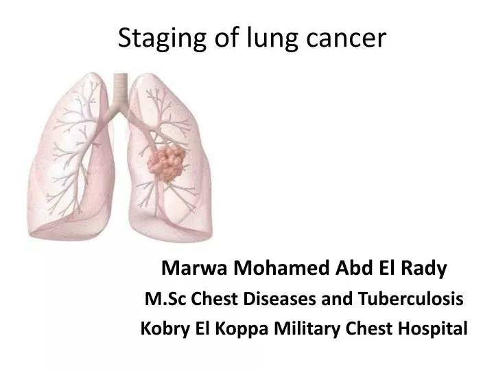
Staging of lung cancer Marwa Mohamed Abd El Rady M.Sc Chest Diseases and Tuberculosis Kobry El Koppa Military Chest Hospital
Introduction • Definition of the stage is an essential part of the approach to patients with cancer, and it has led to the development of a universally accepted stage classification systems for most tumors.
The Union Internationale Contre le Cancer (UICC) and the American Joint Committee on Cancer (AJCC) serve as the official bodies that define, periodically review, and refine the stage classification systems.
What? Staging is a way of describing a cancer, such as where it is located, if or where it has spread, and whether it is affecting the functions of other organs in the body. Why? • Decide what kind of treatment is best. • Predict a patient’s prognosis.
TNM Staging System • Historical background: • Dr. Clifton Mountain that was adopted by the AJCC in 1973 and by the UICC in 1974. • Database of 2,155 patients from the MD Anderson Cancer Center in Houston, TX. • 5,319 cases at the time the lung cancer staging system was last revised in 1997.
A proposal to develop an international effort to inform a future revision of the TNM staging classification for lung cancer originated in 1996. • Data elements and definitions were finalized in October 2002. Data were collected from multiple sources and sites around the globe.
The proposed staging recommendations were presented to the AJCC and UICCin2007and published in the 7th edition of the UICC Staging Manual in 2009. • At the time the database had been submitted from 45 sources in 20 countries. The final data set involved 81,015 cases after exclusion of ineligible cases.
Fundamentals of the Staging System • The T descriptor: defines the extent of the primary tumor. • The N descriptor: the extent of involvement of regional lymph nodes. • The M descriptor: the extent of spread to distant sites.
Definitions for T, N, M DescriptorsT Descriptor • T Primary tumor • T0No primary tumor • T1 Tumor ≤ 3 cm,† surrounded by lung or visceral pleura, not more proximal than the lobar bronchus • T1aTumor ≤ 2 cm† • T1b Tumor> 2 but ≤ 3 cm† • T2 Tumor > 3 but ≤ 7 cm† or tumor with any of the following:Invades visceral pleura, involves main bronchus > 2 cm distal to the carina, atelectasis/obstructive pneumonia extending to hilum but not involving the entire lung. • T2a Tumor > 3 but ≤ 5 cm† • T2b Tumor > 5 but ≤ 7 cm† • T3 Tumor > 7 cm; or directly invading chest wall, diaphragm, phrenic nerve, mediastinal pleura, or parietal pericardium;or tumor in the main bronchus < 2 cm distal to the carina§; or atelectasis/obstructive pneumonitis of entire lung; or separate tumor nodules in the same lobe . • T4Tumor of any size with invasion of heart, great vessels, trachea, recurrent laryngeal nerve, esophagus, vertebral body, or carina;or separate tumor nodules in a different ipsilateral lobe
N Descriptor • NRegional lymph nodes • N0 No regional node metastasis. • N1 Metastasis in ipsilateralperibronchial and/or perihilar lymph nodes and intrapulmonary nodes, including involvement by direct extension. • N2 Metastasis in ipsilateralmediastinal and/or subcarinal lymph nodes. • N3 Metastasis in contralateralmediastinal, contralateralhilar, ipsilateral or contralateral scalene, or supraclavicular lymph nodes.
M Descriptor • MDistant metastasis • M0 No distant metastasis. • M1a Separate tumor nodules in a contralateral lobe; or tumor with pleural nodules or malignant pleural dissemination . • M1b Distant metastasis.
Special situations • TX, NX, MX T, N, or M status not able to be assessed. • Tis Focus of in situ cancer. • T1§ Superficial spreading tumor of any size but confined to the wall of the trachea or mainstem bronchus.
T Descriptor • The size threshold of 3 cm was confirmed as a significant cutpoint and retained as a definition of a T1 vs a T2 tumor. • Significant cutpoints were identified at 2, 5, and 7cm. • The 2-cm and 5-cm additional cutpoints were addressed by the definition of subgroups of T1 (T1a and T1b) and T2 (T2a and T2b).
Patients with additional satellite nodules in the same lobe as the primary tumor had relatively good survival, similar to T3 patients, and therefore are now classified as T3 (previously classified as T4). • An ipsilateral nodule in a different lobe is now classified as T4(previously classified as M1).
Clinically staged patients with pleural dissemination clearly have statistically significant worse survival than T4 by invasion (T4Inv) or than patients with an additional nodule in a different ipsilateral lobe (T4Ipsi Nod).Therefore, patients with pleural dissemination are classified as M1a.
N Descriptor • No changes were made in the N descriptor as defined in the 6th edition of the staging manual. • There was no difference in survival between patients with involvement of only peripheral N1 nodes or hilar N1 nodes,and no difference based on which N2 nodal stations were involved.
The number of involved nodal zones did appear to have a prognostic impact. • The prognostic impact of the number of nodal zones involved could not be confirmed within T stage categories.
M Descriptor • The prognostic impact of one vs more than one contralateral pulmonary nodule could not be assessed. • Slightly worse survival was seen in patients with multiple vs a solitary distant metastasis.
Staging of SCLC • The two stage system originally introduced by the Veterans' Affairs Lung Study Group (VALSG) is widely utilized in staging of SCLC. • Limited disease: is defined as disease confined to the ipsilateralhemithorax and within a single radiotherapy port (corresponding in part to TNM stages I through IIIB). • Extensive disease: is defined as evident metastatic disease outside the ipsilateralhemithorax.
Clinical-diagnostic stage • Medical history. • Physical examination. • laboratory testing. • Radiologic testing. • Tissue sampling.
History and physical examination • Identify symptoms or physical findings suggestive of locally advanced or metastatic disease. • Assess pulmonary health status. • Identify significant comorbidities. • Assess overall health status.
Value: • Each impacts the therapeutic options. • The patient's ability to tolerate treatment. • The disease course independent of disease stage.
History and physical examination • History • Symptoms&Signs: • cough, dyspnea, hemoptysis, postobstructive pneumonia. • Systemic symptoms. • Bone pain . • Dysphagia. • Neurologic abnormalities .
Horner's syndrome. • Hoarseness of voice. • Superior vena cava syndrome. • Pericardial tamponade. • Enlarged supraclavicular and scalene lymph nodes. • Hepatomegaly. • Paraneoplastic syndromes.
Clinical-diagnostic stage • Medical history. • Physical examination. • laboratory testing. • Radiologic testing. • Tissue sampling.
Laboratory tests • Complete blood count. • Serum electrolytes. • Calcium. • (ALT)& (AST). • Alkaline phosphatase. • Creatinine. • Tumor markers.
Clinical-diagnostic stage • Medical history. • Physical examination. • laboratory testing. • Radiologic testing. • Tissue sampling.
Radiographic imaging • Contrast-enhanced CT scanning: • It can characterize the primary tumor. • Define its relationship to the chest wall and mediastinal structures. • It can identify enlarged mediastinal lymph nodes. • Assess contralateral lung, chest wall, or upper abdominal lesions that are suspicious for metastasis.
Radiographic imaging • PET scanning.
Radiographic imaging • Brain CT scanning. • Magnetic resonance imaging (MRI). • Bone scan.