Download
1 / 74
740 likes | 925 Views
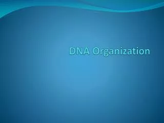
Organization of DNA. Large DNA molecules must be packaged to fit inside the cell and still be functional ! Supercoiling

E N D
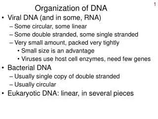
Large DNA molecules must be packaged to fit inside the cell and still be functional ! Supercoiling • mt DNA and the DNA of most prokaryotes are closed circular structures. These molecules may exist as relaxed circles or as supercoiled structures in which the helix is twisted around itself in three-dimensional space. • Supercoiling results from strain on the molecule caused by under- or overwinding the double helix:
Negatively supercoiled DNA: if the DNA is wound more loosely than in Watson-Crick DNA. This form is required for most biologic reactions. • Positively supercoiled DNA: if the DNA is wound more tightly than in Watson-Crick DNA. • Topoisomerases • can change the amount of supercoiling. • make transient breaks in DNA strands by alternately breaking and resealing the sugar-phosphate backbone. • E.g., in Escherichia coli, DNA gyrase (DNA topoisomerase II) can introduce negative supercoiling into DNA, whereas DNA topoisomerase I can relax the supercoils
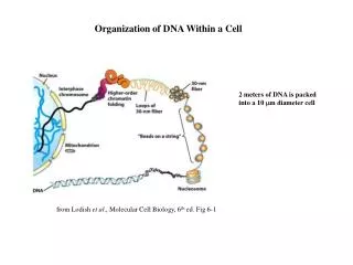
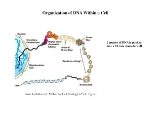
Nucleosomes and Chromatin • Nuclear DNA in eukaryotes is found in chromatin associated with histones and nonhistone proteins. • The basic packaging unit of chromatin is the nucleosome • Histones are rich in lys and arg a positive charge. • Two copies each of histones H2A, H2B, H3, and H4 aggregate to form the histone octamer. • DNA is wound around the outside of this octamer to form a nucleosome (a series of nucleosomes is sometimes called "beads on a string").
Chromosome structure (Nucleofilament)
Organization of human DNA, illustrating the structure of nucleosomes.
Histone H1 is associated with the linker DNA found between nucleosomes to help package them into a solenoid-like structure, which is a thick 30-nm fiber. • Further condensation occurs to eventually form the chromosome. Each eukaryotic chromosome contains one linear molecule of DNA.
Electron micrograph of a protein-depleted human metaphase chromosome, showing the residual chromosome scaffold and loops of DNA. Individual DNA fibers can be best seen at the edge of the DNA loops. Bar = 2μ.(From Paulson JR, Laemmli UK [1977] The structure of histone-depleted metaphase chromosomes. Cell 12:817–828.
Cells in interphase contain two types of chromatin: • Euchromatin is loosely packaged and transcriptionally active. • Heterochromatin is tightly packaged and inactive. • Euchromatin generally corresponds to looped 30-nm fibers. • Heterochromatin is more highly condensed.
Gene expression requires that chromatin be opened for access by transcription complexes (RNA polymerase and transcription factors). • Chromatin-modifying activities include: • Histone acetylation • Histone phosphorylation • During mitosis, all the DNA is highly condensed to allow separation of the sister chromatids. This is the only time in the cell cycle when the chromosome structure is visible. • Chromosome abnormalities may be assessed on mitotic chromosomes by karyotype analysis (metaphase chromosomes) and by banding techniques (metaphase, prophase or prometaphase), which identify aneuploidy, translocations, deletions, inversions, and duplications.
1. Cytosine arabinoside (araC) is used as an effective chemotherapeutic agent for cancer, although resistance to this drug may eventually develop. In certain cases, resistance is related to an increase in the enzyme cytidine deaminase in the tumor cells. This enzyme would inactivate araC to form A. cytosine B. cytidylic acid C. thymidine arabinoside D. uracil arabinoside E. cytidine
2. A double-stranded RNA genome isolated from a virus in the stool of a child with gastroenteritis was found to contain 15% uracil. What is the percentage of guanine in this genome? A. 15 B. 25 C. 35 D. 75 E. 85
3. Endonuclease activation and chromatin fragmentation are characteristic features of eukaryotic cell death by apoptosis. Which of the following chromosome structures would most likely be degraded first in an apoptotic cell? A. Barr body B. 10nm fiber C. 30 nm fiber D. Centromere E. Heterochromatin
DNA Replication and Repair OVERVIEW OF DNA REPLICATION • Genetic information is transmitted from parent to progeny by replication of parental DNA • During DNA replication, the two complementary strands of parental DNA are pulled apart. Each of these parental strands is then used as a template for the synthesis of a new complementary strand (i.e., semiconservative). • During cell division, each daughter cell receives one of the two identical DNA molecules.
The bacterial chromosome is a closed, double-stranded circular DNA molecule having a single origin of replication. • Separation of the two parental strands of DNA creates two replication forks that move away from each other in opposite directions • Replication is, thus, a bidirectional process. • The two replication forks eventually meet, resulting in the production of two identical circular molecules of DNA.
Each eukaryotic chromosome contains one linear molecule of DNA having multiple origins of replication. • Bidirectional replication occurs at each origin. • Completion of the process results in the production of two identical linear molecules of DNA. • DNA replication occurs in the nucleus during the S phase of the eukaryotic cell cycle. • The two identical sister chromatids are separated during mitosis.
COMPARISON OF DNA AND RNA SYNTHESIS • The overall process of DNA replication requires the synthesis of both DNA and RNA. • These two types of nucleic acids are synthesized by DNA polymerases and RNA polymerases
3'→5'-Exonuclease activity enables DNA polymerase III to “proofread” the newly synthesized DNA strand.
Polymerases and Nucleases • Polymerases are enzymes that synthesize nucleic acids by forming phosphodiester (PDE) bonds. Nucleases are enzymes that hydrolyze PDE bonds • Exonucleases remove nucleotides from either the 5' or the 3' end of a nucleic acid. • Endonucleases cut within the nucleic acid and release nucleic acid fragments.
Table 1-2-1. Comparison of DNA and RNA Polymerases *Certain DNA and RNA polymerases require RNA templates. These enzymes are most commonly associated with viruses.
Similarities include: • The newly synthesized strand is made in the 5'3' direction. • The template strand is scanned in the 3'5' direction. • The newly synthesized strand is complementary and antiparallel to the template strand. • Each new nucleotide is added when the 3' hydroxyl group of the growing strand reacts with a nucleoside triphosphate, which is base-paired with the template strand. Pyrophosphate (PPi, the last two phosphates) is released during this reaction.
Differences include: • The substrates for DNA synthesis are the dNTPs, whereas the substrates for RNA synthesis are the NTPs. • DNA contains thymine, whereas RNA contains uracil. • DNA polymerases require a primer, whereas RNA polymerases do not. That is, DNA polymerases cannot initiate strand synthesis, whereas RNA polymerases can. • DNA polymerases can correct mistakes ("proofreading"), whereas RNA polymerases cannot. DNA polymerases have 3'5' exonuclease activity for proofreading.
Steps of DNA Replication in E. coli 1. The base sequence at the origin of replication is recognized and bound by the dna A protein. The two parental strands of DNA are pulled apart to form a "replication bubble". 2. Helicase uses energy from ATP to break the hydrogen bonds holding the base pairs together. This allows the two parental strands of DNA to begin unwinding and forms two replication forks. 3. Single-stranded DNA binding protein (SSB) binds to the single-stranded portion of each DNA strand, preventing the strands from reassociating and protecting them from degradation by nucleases.
4. Primase synthesizes a short (about 10 nucleotides) RNA primer in the 5'3' direction, beginning at the origin on each parental strand. The parental strand is used as a template for this process. RNA primers are required because DNA polymerases are unable to initiate synthesis of DNA, but can only extend a strand from the 3' end of a preformed "primer".
5. DNA polymerase III begins synthesizing DNA in the 5'3' direction, beginning at the 3' end of each RNA primer. The newly synthesized strand is complementary and antiparallel to the parental strand used as a template. This strand can be made continuously in one long piece and is known as the "leading strand". • The "lagging strand" is synthesized discontinuously as a series of small fragments (about 1,000 nucleotides long) known as Okazaki fragments. Each Okazaki fragment is initiated by the synthesis of an RNA primer by primase, and then completed by the synthesis of DNA using DNA polymerase III. Each fragment is made in the 5'3' direction. • There is a leading and a lagging strand for each of the two replication forks on the chromosome.
6. RNA primers are removed by DNA polymerase I. This enzyme removes the ribonucleotides one at a time from the 5' end of the primer (5'3' exonuclease). DNA polymerase I also fills in the resulting gaps by synthesizing DNA, beginning at the 3' end of the neighboring Okazaki fragment. 7. Both DNA polymerase I and III have the ability to "proofread" their work by means of a 3'5' exonuclease activity. If DNA polymerase makes a mistake during DNA synthesis, the resulting unpaired base at the 3' end of the growing strand is removed before synthesis continues.
Removal of RNA primer and filling of the resulting “gaps” by DNA polymerase-I
8. DNA ligase seals the "nicks" between Okazaki fragments, converting them to a continuous strand of DNA. • DNA gyrase (DNA topoisomerase II) provides a "swivel" in front of each replication fork. As helicase unwinds the DNA at the replication forks, the DNA ahead of it becomes overwound and positive supercoils form. DNA gyrase inserts negative supercoils by nicking both strands of DNA, passing the DNA strands through the nick, and then resealing both strands again. • DNA topoisomerase I can relieve supercoiling in DNA molecules by the transient breaking and resealing of just one of the strands of DNA. • Replication is completed when the two replication forks meet each other on the side of the circle opposite the origin.
Quinolones are a family of drugs that block the action of topoisomerases. Nalidixic acid kills bacteria by inhibiting DNA gyrase. • Inhibitors of eukaryotic topoisomerase II (etoposide, teniposide) are becoming useful as anticancer agents.
9. The mechanism of replication in eukaryotes is believed to be very similar to this. However, the details have not yet been completely worked out.
Eukaryotic DNA Polymerases • DNA polymerase δ elongates leading and Okazaki fragments during replication. • DNA polymerase α initiates synthesis and has primase activity. • DNA polymerase γ replicates mitochondrial DNA. • DNA polymerases β and ε are thought to participate primarily in DNA repair. DNA polymerase ε may substitute for DNA polymerase δ in certain cases.
Telomerase • Telomeres are repetitive sequences at the ends of linear DNA molecules in eukaryotic chromosomes. • With each round of replication in most normal cells, the telomeres are shortened because DNA polymerase cannot complete synthesis of the 5' end of each strand. • This contributes to the aging of cells, because eventually the telomeres become so short that the chromosomes cannot function properly and the cells die.
Telomerase is an enzyme in eukaryotes used to maintain the telomeres. It contains a short RNA template complementary to the DNA telomere sequence, as well as telomerase reverse transcriptase activity (hTRT). Telomerase is thus able to replace telomere sequences that would otherwise be lost during replication. Normally telomerase activity is present only in embryonic cells, germ (reproductive) cells, and stem cells, but not in somatic cells. • Cancer cells often have relatively high levels of telomerase, preventing the telomeres from becoming shortened and contributing to the immortality of malignant cells.
Telomere Ends are Loops Not “naked” ends • Telomeres and TRF1 (telomere repeat-binding factor) protein stabilize ends for two reasons (replication gaps & DNase degradation) • Protection against recombination and NHEJ by distinguishing them from DSBs
Reverse Transcriptase • Reverse transcriptase is an RNA-dependent DNA polymerase that requires an RNA template to direct the synthesis of new DNA. Retroviruses, most notably HIV, use this enzyme to replicate their RNA genomes. • DNA synthesis by reverse transcriptase in retroviruses can be inhibited by AZT, ddC, and ddI.
Eukaryotic cells also contain reverse transcriptase activity. • Associated with telomerase (hTRT). • Encoded by retrotransposons (residual viral genomes permanently maintained in human DNA) that play a role in amplifying certain repetitive sequences in DNA
Bridge to Pharmacology • Quinolones and DNA Gyrase: Quinolones and fluoroquinolones inhibit DNA gyrase (prokaryotic topoisomerase II), preventing DNA replication and transcription. These drugs, which are most active against aerobic gram-negative bacteria include: • Nalidixic acid • Ciprofloxacin • Norfloxacin • Resistance to the drugs has developed overtime; current uses include treatment of gonorrhea, and upper and lower urinary tract infections in both sexes.