Download
1 / 20
200 likes | 333 Views
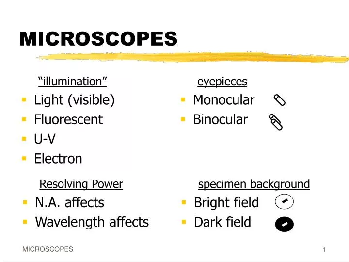
“illumination” Light (visible) Fluorescent U-V Electron. eyepieces Monocular Binocular. specimen background Bright field Dark field. MICROSCOPES. Resolving Power N.A. affects Wavelength affects. PARTS & TERMS. Lenses (ocular, objective) Total Magnification

E N D
“illumination” Light (visible) Fluorescent U-V Electron eyepieces Monocular Binocular specimen background • Bright field • Dark field MICROSCOPES Resolving Power • N.A. affects • Wavelength affects MICROSCOPES
PARTS & TERMS • Lenses (ocular, objective) • Total Magnification • ocular mag. X objective mag. • Condenser • Diaphram MICROSCOPES
PARTS & TERMS • Numerical Aperture (N.A.) optical characteristic of a lens N.A. = i sinq N.A. increases with magnification MICROSCOPES
(l innm) R.P. = wavelength of illumination (2)N.A. PARTS & TERMS • Resolving Power (R.P.) • size of the smallest discernable detail • minimum distance between objects so that they are able to be distinguished as separate [ RP of human eye ~ 0.2mm = ______m ] MICROSCOPES
l = 650nm, NA = 1.25 R.P. = l/2NA = _____nm = _____ m l = 450nm, NA = 1.25 R.P. = l/2NA = _____nm = _____ m Resolving Power (R.P.) l = 650nm, NA = 0.25 R.P. = l/2NA = _____nm = _____ m 1300 1.3 260 0.260 180 0.180 MICROSCOPES
Resolving Power (R.P.) R.P. = l/2NA Larger or Smaller R.P. = BETTER ? • R.P. improves as N.A. increases or decreases? • R.P. improves as l increases or decreases? MICROSCOPES
Types of Scopes MICROSCOPES
Types of Scopes MICROSCOPES
Types of Scopes Compound Light • Bright or Dark field • Use blue light or blue filter for shorter l • R.P. ~ 0.2m MICROSCOPES
Types of Scopes Fluorescent • Dark field compound light microscope • Uses U-V for side illumination of specimen, fluorescent parts or dyes give off visible light that is viewed • R.P. ~ 0.2m MICROSCOPES
Types of Scopes UltraViolet (U-V) • Uses U-V as illumination (shorter l) • Image recorded then viewed… still or video camera image(should NOT look directly at U-V!) • Special lens material (quartz),glass absorbs U-V • R.P. ~ 0.15m MICROSCOPES
Types of Scopes Electron Microscopes • Use electron beam for illumination, magnets for “lenses”, video to view • Transmission Electron Microscope (TEM) • beam passes thru, view internal structure • Scanning Electron Microscope (SEM) • beam reflects, see external structure, texture MICROSCOPES
Types of Scopes Transmission Electron Microscope (TEM) MICROSCOPES
Types of Scopes Transmission Electron Microscope (TEM) • Special specimen preparation (vacuum) • Working magnification: whole bacteria….. 8,000x - 10,000x thin section, viruses…. 30,000x - 40,000x • Maximum magnification ~100,000x • R.P. ~0.001m = ______nm MICROSCOPES
rickettsia herpes simplex
Types of Scopes Scanning Electron Microscope (SEM) MICROSCOPES
Types of Scopes Scanning Electron Microscope (SEM) • Special specimen preparation(vacuum & conductor) • Working magnification: 14,000x - 50,000x • Maximum magnification ~130,000x • R.P. ~0.01m = ______nm MICROSCOPES
Q fever (rickettsia) Image enhancement