Download
1 / 13
130 likes | 288 Views
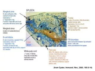
(from Cyster, Immunol. Rev., 2003. 195:5-14). White pulp nodule within spleen. (from Fu and Chaplin, Annu. Rev. Immunol. 1999. 17:399-433). Follicular dendritic cells (FDC)

E N D
White pulp nodule within spleen (from Fu and Chaplin, Annu. Rev. Immunol. 1999. 17:399-433)
Follicular dendritic cells (FDC) - abundant complement receptors and Fc receptors - focus immune complexes within the B cell follicle - crucial for the development of effective isotype-switched and memory B cell responses. FDC precursors are radiation-resistant Dendritic cells and lymphocytes are from radiation-sensitive bone marrow-derived precursors Development of GC structures (PNA+) depends on intercellular signaling via CD40 and CD40L, CD19, CD28, and B7-2, among others. In the absence of T cells, spleens have no GC’s, but do have white pulp nodules containing NK, B and DC, and FDC clusters Mesenteric LN are different - can have ‘GC’s’ without FDC clusters
TNF, LT (or LTa) and LTß (LT1aß2) are structurally related and encoded within same 25 kb portion of MHC III • TNFRI and TNFRII broadly-expressed • LTßR expressed on stromal cells in various tissues • LTß-mediated responses involve cell-cell contact (from Fu and Chaplin, Annu. Rev. Immunol. 1999. 17:399-433)
LT-/- mice: Loss of most lymph nodes, and Peyer’s Patches. Single mesenteric LN’s in 2-4% of mice. Spleens show loss of discrete T and B cell zones, loss of marginal sinus MAdCAM-1–staining, loss of discrete B cell follicles and FDC networks, and loss of (PNA+) GC B cell clusters Impaired specific Ig responses and affinity maturation TNFRI-/- and TNF-/- mice: No loss of lymph nodes Spleens show absence of marginal sinus MAdCAM-1 staining, lack of discrete B cell follicles and FDC networks, and lack of splenic GC. But - segregation of T and B cell zones retained
LTßR +/+ India ink draining from footpad identifies popliteal lymph node LTßR -/- No popliteal lymph node (from Fütterer et al, Immunity1998;9:59-70)
Bone marrow chimera experiments (eg. LTa-/- BM -> irradiated WT recipient, generates mice with segregated T/B zones, but no FDC’s) show that: The ability to form discrete white pulp B cell and T cell zones is a fixed feature of the microenvironment, imprinted by the time mice reach maturity. Whereas - B cell follicle structure (FDC clusters) is dependent on continued presence of LTa- or LTß-expressing cells TNF- or TNFRI-deficient mice also lack FDC clusters Indirect evidence that FDC response to LTß and TNF is required: Radiation-resistant LTßR- and TNFRI-expressing cells are required to generate FDC clusters in bone marrow chimeras
Irradiation chimeras (previous slide) also showed that: LT-expressing cells that are required for formation and maintenance of FDC network are bone marrow-derived. B cell MiceLymphocytesFolliclesFDCMadCAM-1 WT + + + + RAG-/- NK only - - +/- BCR-/- NK, T - - +/- TCR-/- B, NK + + + CD3e B + + + transgenic Conclusion: LT- and TNF-expressing cells that are required for formation and maintenance of FDC network in spleen are B cells (from Fu and Chaplin, Annu. Rev. Immunol. 1999. 17:399-433)
Mem-TNF Tg mice have normal LN development • Mice without secreted TNF but with functional normally-regulated and expressed membrane-bound TNF (Mem-TNF∆/∆ mice) were created by knocking-in the uncleavable ∆1-9,K11E TNF allele. • In contrast to TNF-deficient mice (TNF-/-), mem-TNF supported many features of lymphoid structure, except generation of primary B cell follicles. • Splenic chemokine production was nearly normal in Mem-TNF mice • Mem-TNF was suboptimal for development of inflammation. Ruuls et al, 2001, Immunity 15:533
Site-directed mutagenesis of the TACE cleavage site in exons II and III - removal of amino acids 1-9 and K11->E11 mutation. Homologous recombination to ES cells, injection into blastocysts, breeding chimeric animals -> +/- X +/- -> ∆/∆ Mem-TNF mice A: LPS-induced TNF in cell supernatants of peritoneal-exudate cells (PEC’s) B: FACS-staining for surface TNF on LPS-stimulated PEC’s C: TNF RNA in LPS-stimulated PEC’s Not shown - expression of LTa and LTß was unaffected Ruuls et al, 2001, Immunity 15:533
SPLEEN No primary B cell follicles T:B segregation less than in WT, but improved over TNF-/- Marginal zone metallophilic macrophages restored MadCAM restored PNA+ Germinal centres CR1+ FDC networks Ruuls et al, 2001, Immunity 15:533
Peyer’s Patch organization restored Lymph Nodes - similar to spleen Ruuls et al, 2001, Immunity 15:533
Rescue of chemokine expression in spleen of mem-TNF ∆/∆ mice Ruuls et al, 2001, Immunity 15:533