Download
1 / 18
190 likes | 359 Views
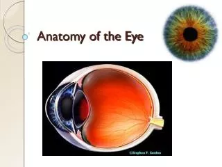
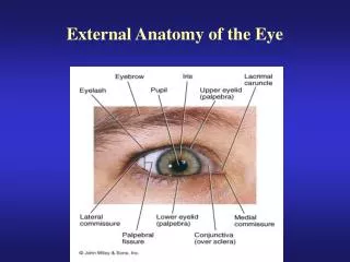
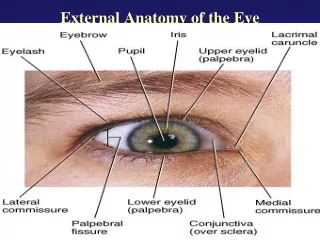
Lecture 1: Eye Anatomy. Liana Al-Labadi, O.D. Eye Anatomy. Eye Anatomy. The orbital bone The eye socket Formed by: Cheekbone Forehead Temple Side of nose Eye is cushioned within orbit by pads of fat Lacrimal gland Produces tears Tears drain through the nasolacrimal duct.

E N D
Lecture 1: Eye Anatomy Liana Al-Labadi, O.D.
Eye Anatomy http://everlastingelephants.blogspot.com/2009/08/what-is-eye-cataract.html
Eye Anatomy • The orbital bone • The eye socket • Formed by: • Cheekbone • Forehead • Temple • Side of nose • Eye is cushioned within orbit by pads of fat • Lacrimal gland • Produces tears • Tears drain through the nasolacrimal duct http://commons.wikimedia.org/wiki/File:Eye_orbit_anatomy_anterior2.jpg http://mwsu-bio101.ning.com/forum/topics/distinct-human-celltypes-1?commentId=2263214%3AComment%3A10331
Eye Anatomy • Eyelids (L): • Protection: • Protects eye from foreign matter (dust, dirt, debris) • Protects against bright light that might damage the eye • Help spread tears over surface of eye- moist & comfort • Eyelashes (L): • Filter out foreign matter • prevent it from getting into eye http://www.medical-look.com/human_anatomy/organs/Eyelids_and_eyelashes.html
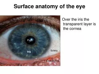
Eye Anatomy • Conjunctiva (Conj): • Thin, clear layer of skin • Covering of the front of eye • Covers the sclera and the inside of the eyelids • Function: • Keeps bacteria and foreign material from getting behind eye http://www.images.missionforvisionusa.org/anatomy/2005/11/conjunctiva-answers.html
Eye Anatomy • Sclera (S): • “White of the eye” • Tough, opaque tissue that extends around the eye • Surrounds the eye and gives the eye its shape • The sclera is attached to the extraocular muscles http://www.thirdeyehealth.com/sclera.html
Eye Anatomy • Extraocular Muscles • 6 extraocular muscles that are attached to each eye • Help move the eye left, right, up, down and diagonally • These 6 muscles are: • Superior rectus • Inferior rectus • Medial rectus • Lateral rectus • Inferior oblique • Superior oblique http://media.photobucket.com/image/introduction%20to%20eye%20anatomy/trimurtulu/Eye.jpg
Eye Anatomy • Cornea (K): • Clear layer at the front & center of eye • Located in front of the iris (colored part of eye) • Function: • Focus light as it enters eye • Avascular • Only organ that has no blood vessels http://commons.wikimedia.org/wiki/File:Cornea.jpg
Eye Anatomy • Anterior Chamber (AC): • Fluid-filled space • Behind the cornea & in front of the iris • Fluid = Aqueous humor (AH) • AH helps nourish the cornea & the lens http://www.djo.harvard.edu/files/2528_310.jpg • http://www.goodhope.org.uk/departments/eyedept/angleclosureetc.htm
Eye Anatomy • Pupil (P): • Central opening of iris • Iris (I): • Ring shaped tissue • Colored part of eye • Controls the amount of light that enters the eye • Two muscle fibers: • Contraction • Constricts pupil in bright light • Dilation • Dilates pupil in dark http://www.bioconsulting.com/Bio_Tech_Assessment.html • http://www.goodhope.org.uk/departments/eyedept/angleclosureetc.htm
Eye Anatomy • Anterior Chamber Angle • Located where the cornea meets the iris • Trabecular Meshwork • Site where aqueous humor drains out of eye • If AH cannot properly drain out • Pressure build up inside eye • Causes optic nerve damage & evetually vision loss = glaucoma http://seniorhealth.about.com/library/conditions/blglaucoma2.htm
Eye Anatomy • Posterior Chamber (PC): • Fluid-filled space • Aqueous Humor! • Immediately behind the iris but infront of the lens http://seniorhealth.about.com/library/conditions/blglaucoma2.htm
Eye Anatomy • Crystalline Lens: • Clear, flexible structure • Behind the iris & pupil • Surrounded by a ring of muscular tissue – ciliary body • The lens & ciliary body help control fine focusing of light as it passes through the eye http://www.smartplanet.com/business/blog/smart-takes/artificial-lens-implant-to-give-patients-high-definition-vision-better-than-2020/2558/
Eye Anatomy • Vitreous Chamber: • Located behind the lens & in front of the retina • Filled with a gel-like fluid called the vitreous humor • The vitreous help maintain the shape of the eye http://www.ophthobook.com/questions/question-how-many-chambers-are-there-in-the-eye
Eye Anatomy • Retina: • Acts like the film in a camera to create an image • Consists of a specialized layer of cells • Converts light signals into nerve signal then send these signals to the optic nerve • Optic nerve carries the signals to the brain • The brain helps process the image • Rods- low light situations • Cones- allows you to see color hhttp://www1.appstate.edu/~kms/classes/psy3203/EyePhysio/human_retina.htm http://www.answersingenesis.org/tj/v13/i1/retina.asp
Eye Anatomy • Macula • Located in the central part of the retina • Responsible for giving sharp central vision • Used for reading, recognizing faces, and watching TV • Any disease that affects the macula will cause a change & impairment in the central vision http://www.dukehealth.org/eye_center/specialties/macular_degeneration/care_guides/macular_degeneration_frequently_asked_questions
Eye Anatomy • Choroid • A layer of tissue that is: • Located under the retina • Separates retina & sclera • Mostly made up of blood vessels • Helps nourish the retina by carrying the blood supply to the eye’s internal structures http://www.cnib.ca/en/your-eyes/eye-conditions/amd/the-eye/basics/Default.aspx
Eye Anatomy • Optic Nerve • A bundle of 1 million nerve fibers • Responsible for transmitting nerve signals from the eye to the brain • The optic disc is the front surface of the optic nerve • The optic disc is visible on the retina http://cssd.us/body.cfm?id=802 http://www.wollongong.youronlinecommunity.com.au/wollongong-online/2008/50/walkthrulife/eye-health.html