Eye Anatomy
370 likes | 1.58k Views
Eye Anatomy. Orthopedic Assessment III – Head, Spine, and Trunk with Lab PET 5609C. Clinical Anatomy. Orbit: Cavity or socket of the skull which houses the eye Protects and stabilizes the eye Serves as attachment site for extrinsic muscles

Eye Anatomy
E N D
Presentation Transcript
Eye Anatomy Orthopedic Assessment III – Head, Spine, and Trunk with Lab PET 5609C
Clinical Anatomy • Orbit: • Cavity or socket of the skull which houses the eye • Protects and stabilizes the eye • Serves as attachment site for extrinsic muscles • Orbital Margins – bases which open in the face (4 borders) • Supraorbital margin – frontal bone • Inraorbital margin – zygomatic and maxilla bones • Lateral margin – zygomatic and frontal bones
Yellow – Frontal Bone Blue – Zygomatic Bone Purple – Maxilla Bone Clinical Anatomy
Clinical Anatomy • Orbital Anatomy: • Anterior aspect or roof • Frontal Bone • Posterior aspect • Sphenoid Bone • Medial aspect • Lacrimal, ethmoid, maxillary, and sphenoid bones • Lateral aspect • Zygomatic and sphenoid bones • Orbit is thickest
Clinical Anatomy Frontal Bone Ethmoid Bone Lacrimal Bone Sphenoid Bone Zygomatic Bone Maxilla Bone
Superior Orbital Fissure Opening between lesser and greater wings of sphenoid bone Allows cranial nerves, arteries, and veins to communicate with eye Optic Canal Foramen which the optic nerve passes to reach the brain Optic Nerve Cranial nerve II Transmits visual information from the retina to the brain Clinical Anatomy
Clinical Anatomy Optic Fissure Superior Orbital Fissure
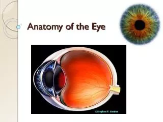
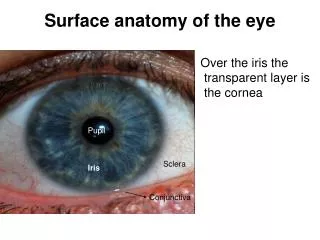
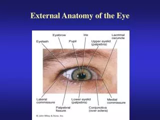
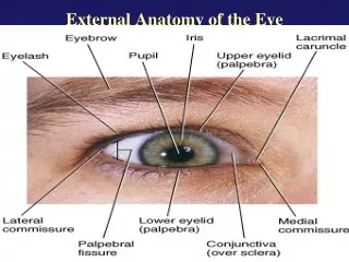
Sclera: White of the eye Tough, opaque tissue that serves as the eye's protective outer Optic nerve is attached to the sclera at the very back of the eye Pupil: Opening in center of iris Size of the pupil determines the amount of light that enters the eye Pupil size is controlled by the dilator and sphincter muscles of the iris Neurological Function – pupils reaction to light Clinical Anatomy
Iris: Colored part of the eye Controls light levels inside the eye Divides the anterior chamber from posterior chamber Color comes from microscopic pigment cells (melanin) The color, texture, and patterns of each person's iris are as unique as a fingerprint Muscles acting on Iris: Sphincter muscle: In bright light, the sphincter contracts, causing the pupil to constrict Dilator muscle: Dilates the eye in dim lighting Clinical Anatomy
Conjunctiva: Thin mucous membrane that covers the outer surface of the eye (sclera) Lines inside of the eyelids Anteriorly - continous with the cornea Nourished by tiny blood vessels (nearly invisible to the naked eye) Secretes oils and mucous that moisten and lubricate the eye Clinical Anatomy
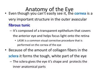
Cornea: Transparent, dome-shaped window covering the front of the eye (normally clear with a shiny surface) Powerful refracting surface (provides 2/3 of the eye's focusing power) Extremely sensitive More nerve endings in the cornea than anywhere else in the body Clinical Anatomy
Ciliary Body: Lies behind the iris Attached to the ciliary body are tiny fiber ligaments (zonules) – suspend the lens Produces aqueous humor (clear fluid that fills the front of the eye) Controls accommodation to light by changing the shape of the lens Ciliary body contracts - zonules relax and lens thicken, ↑ the eye's ability to focus up close Ciliay body relaxes - zonules contract and lens becomes thinner, adjusting the eye's focus for distance vision Lens: Located just behind the iris Focuses light onto the retina Clinical Anatomy
Retina: Multi-layered sensory tissue that lines the back of the eye Contain millions of photoreceptors that capture light rays and converts them into electrical impulses Impulses: Optic nerve to Brain (images) Cones (6 million) Bright light (help us differentiate color) Rods (125 million) Peripheral and night vision Clinical Anatomy
Blink Reflex • Corneal Reflex - Blink Reflex • Involuntary blinking of the eyelids elicited by stimulation (touching or a foreign body) of the cornea, or bright light • Should elicit response of the opposite eye also • Time = 0.1 second • Purpose - protect the eyes from foreign bodies and bright lights • Controlled by: • Cranial nerve V (trigeminal nerve) - senses the stimulus on the cornea, lid, or conjunctiva. • Cranial nerve VII (facial nerve) – initiates motor response • Use of contact lenses may diminish or abolish this reflex
Muscular Anatomy: Inferior Rectus Superior Rectus Medial Rectus Lateral Rectus Inferior Oblique Superior Oblique Clinical Anatomy
Clinical Anatomy • Eye Movement Terminology: • Duction – movement of one eye by itself • Version – movement of the 2 eyes in the same direction • Adduction – eye looks toward the nose • Abduction – eye looks toward the ear • Dextroversion – both eyes look to the right • Levoversion – both eyes look to the left • Supraversion – both eyes upgaze • Infraversion - downgaze
Medial Rectus: Strongest of the extra-ocular muscles Most mass of EOMs Most anterior insertion (extra leverage) Action – Adduction (eyes move towards the nose) Lateral Rectus: Action - Abduction Clinical Anatomy
Superior Rectus: Action – elevation, upward rotation Rotation – angles nasally toward site of origin Tendon of the Superior Oblique muscle passes underneath the SR Clinical Anatomy
Inferior Rectus: Action – depression, downward rotation, adduction Clinical Anatomy
Superior Oblique: Keeps the eyeballs level as the head tilts Longest of the EOMs Passes through a “pully” called the trochlea Redirects the action Action: Abduction of globe Depression of globe Rotation of globe Clinical Anatomy
Inferior Oblique: Passes underneath the inferior rectus Action: Elevation of globe Adduction of globe Rotation of globe Keeps the eyeballs level as the head tilts Clinical Anatomy