Download

1 / 15
160 likes | 338 Views
Device for Collecting Stress Images of Subtalar Joint. Patrick Melton Daniel Escobar 12/2/10. Introduction.

E N D
Device for Collecting Stress Images of Subtalar Joint Patrick Melton Daniel Escobar 12/2/10
Introduction • It is difficult to determine if the subtalar joint is unstable through current methods of examination due to the cost and difficulty of use of the equipment necessary to make reliable measurements. • Producing radiographs to allow for accurate measurements of the subtalar joint for stability analysis has been refined for more practical uses.
Purpose • Create a device that will align the subtalar joint to allow for the best possible radiograph and measurements to determine the stability of the joint.
Cobey View Reilingh, M., Beimers, L., Tuijthof, G., Stufkens, S., Maas, M., & van Dijk, C. (2010). Measuring hindfoot alignment radiographically: the long axial view is more reliable than the hindfoot alignment view. Skeletal Radiology, 39(11), 1103-1108.
Long Axial Alignment View • Most reliable and accurate measurements, highest correlation coefficients. • Reilingh, M., Beimers, L., Tuijthof, G., Stufkens, S., Maas, M., & van Dijk, C. (2010). Measuring hindfoot alignment radiographically: the long axial view is more reliable than the hindfoot alignment view. Skeletal Radiology, 39(11), 1103-1108.
Current Progress • Weekly meetings with project advisors discussing literature review, conceptual designs, and future objectives. • Literature Review • Subtalar joint anatomy • Methods of radiographing the subtalar joint for best possible measurements. • Telos device – same concept but difficult to use.
Current Progress (cont’d.) • Considering conceptual designs for stress device • Meeting with Portsmouth Naval Surgeon to discuss potential design ideas • Modeling preliminary designs in Solid Works
Design Considerations • Design allows for inversion of the calcaneus for hindfoot assessment in weight bearing position. Price, Mark. (Designer). (2006). Measuring hindfoot with dynastat. [Web]. Retrieved from http://www.gp-training.net/rheum/gait/gait2.jpg
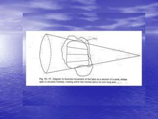
Design Considerations (Ctd) • Applies non-weight bearing inversion stress. • Allows for analysis of subtalar tilt angle. Brantigan, JW, Pedegana, LR, Lippert, FGInstability of the subtalar joint. Diagnosis by stress tomography in three casesJ Bone Joint Surg Am 1977 59: 321-324
Future Objectives • Model the designs in Solid Works and analyze Finite Element model for stresses and factors of safety. • Discuss designs with advisors and make any necessary modifications. • Determine materials needed for constructing device and the cost of the design.