Discovering DNA: A Molecular Masterpiece Unveiled
300 likes | 329 Views
Explore the history and structure of DNA, from Hammerling's 1930s discoveries in algae to Watson & Crick's groundbreaking revelation in 1953. Understand DNA replication through animations and explanations of nucleotide components, hydrogen bonding rules, and DNA shape. Learn about the stability of DNA strands based on base pair bonds and discover the complexities of gene arrangement and coding within DNA. Unravel the intricate process of DNA replication, including strand separation, complementary strand building, and the unique roles of various enzymes involved.

Discovering DNA: A Molecular Masterpiece Unveiled
E N D
Presentation Transcript
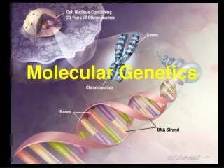
Unit 3 MOLECULAR GENETICS
I] DNA, RNA and PROTEIN SYNTHESIS The Analogy our textbook represents the DNA and there is only one copies. the textbook never leaves the classroom [nucleus] in this textbook all of the left hand pages are identicalto the right hand pages if we wish to send a chapter to the shop we transcribe the chapter that way, if the transcribed chapter is destroyed, we still have the original
A] HISTORY of DNA Hammerling, 1930’s using algae discovered that the nucleus is source of heredity
knew that the nucleus contains proteins and DNA but DNA did not seem complex enough 4 base pairs only
Recall, there are 20 amino acids while proteins did seem complex enough This led to the theory that proteins were the molecules of inheritance.
when they tried to confirm that proteins control heredity using viruses, Hershey & Chase actually showed that DNA controls heredity http://www.youtube.com/watch?v=YG3d77SRWZI
With the help of “borrowed” information from Rosalind Franklin Watson & Crick determined the structure of DNA in 1953
OH HOH2C O OH 1] Parts of a nucleotide a nitrogenous base a phosphate pyrimadine T – thymine C – cytosine O II HO—P—OH I OH a sugar – deoxyribose purine A- adenine G-guanine
O II HO—P—OH I O CH2 O OH combined to form a nucleotide 2] arrangement of nucleotides a] the sugar bonds to the last phosphate at carbon 3 and so sugar end of DNA is called the 3’ end b] the phosphate bonds to the next sugar at carbon5 and so the phosphate end of DNA is called the 5’ end
c] the two strands are arranged as shown below 5’ end l P l sugar—base…… 3’ end l base—sugar l P l base—sugar l P l base—sugar l P l 5’ end l P l sugar—base…… l P l sugar—base…… l 3’ end
3. Hydrogen bonding – adenine forms 2 hydrogen bonds with thymine – cytosine forms 3hydrogen bonds with guanine Since A – T and C – G are the usual bondings, if the amount of A does not equal T or the amount of C does not equal G, then the DNA is not double stranded
Two equal sized pieces of DNA. Which is more stable? Why? G A A T T C T A A T A T C C T T A A G A T T A T A G A G C C G C C T A G C G T C G G C G G A T C G C Sections of the DNA with more C-G bonds, are more stable. [3 H-bonds vs. 2 H-bonds]
4. DNA shape – the stands are arranged in a double helix
5. The letter P represents phosphate; S represents sugar and the letters A, T, C and G are nitrogenous bases P P P P P P P P P P P P P P S S S S S S S S S S S S S S A T A C G C A C T C G A T G T A T G C G T G A G C T A C S S S S S S S S S S S S S S P P P P P P P P P P P P P P 5’ 3’ 3’ 5’
6. Normally this is drawn as shown below T A __A C G C A C T C G A T G T A T G G T G __ _ C T A C C A G
DNA in the cell a. each human cell has about 3 billion base pairs b. these are arranged into about 25,000 genes c. in addition, up to 50% of the DNA does not code for any protein d. in total, the DNA from one cell would be 2m in length e. double stranded DNA [dsDNA] is very stable, but single stranded DNA [ssDNA] is very easily broken
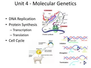
C. DNA Replication 1. Introduction a. before a cell divides it must copy all of its DNA b. it must do this while keeping the amount of ssDNA to a minimum c. also, the DNA will have to be unwound to allow access to the bases
DNA Replication Animations http://www.youtube.com/watch?v=5VefaI0LrgE
1. Separating the strands a] DNA helicase[1] – breaks H-bonds between the strands at an “origin of replication” b] DNA topoisomerase[2] - releases tension by unwinding breaking backbones temporarily c] replication fork[3] - an area where the two strands are separated d] ssB’s [4] - single-stranded binding proteins keep strands from re-annealing
Ignore the Tweed 5’ 3’
2. Building complementary strands a] starting DNA primase[5] - creates a 10-60 base strand of RNA called an RNA-primer [6] at the exposed 3’end of the replication fork [the cell has to use a piece of RNA to start the process because DNA will not stick to DNA as well as RNA will]
3’ 5’ 5’ 3’
b] Building -the enzymeDNA polymerase III[7]- adds complimentary bases that are phosphorylatedmoving along the old DNA in the 3’ to 5’ direction - the energy released by removing 2 Pi’s allows the bonding • the strand that is copied continuously [3’ 5’] strand is • called the leading strand [8]
c] The other strand DNA polymerase III only moves 3’ 5’ - so the lagging strand [9] has to be builtin pieces - theseshortpieces are called Okazaki fragments[10]
3’ 5’ 5’ 3’ 5’ 3’
d] Finishing DNA polymerase I[11] removes the RNA primers & replaces them with the appropriate DNA bases DNA ligase[12] attaches Okazaki fragments by bonding sugar and phosphates
C. DNA REPLICATION 3’ 5’ 5’ 3’ 5’ 3’
3’ 5’ 5’ 3’ 5’ 3’
3’ 5’ 5’ 3’ 5’ 3’