UNIT 3: Molecular Genetics
500 likes | 651 Views
Chapter 5: The Structure and Function of DNA Chapter 6: Gene Expression Chapter 7: Genetic Research and Biotechnology. UNIT 3: Molecular Genetics. UNIT 3. Chapter 5: The Structure and Function of DNA. What are the structures and functions of DNA and RNA?

UNIT 3: Molecular Genetics
E N D
Presentation Transcript
Chapter 5: The Structure and Function of DNA Chapter 6: Gene Expression Chapter 7: Genetic Research and Biotechnology UNIT 3: Molecular Genetics
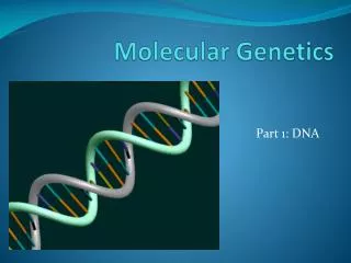
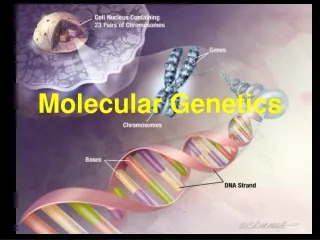
UNIT 3 Chapter 5: The Structure and Function of DNA What are the structures and functions of DNA and RNA? Scientists now know that DNA, shown here in its uncondensed form, carries genetic information that defines many of an organism’s traits (including behaviours) and its predisposition for certain diseases. Chapter 5: The Structure and Function of DNA
UNIT 3 Chapter 5: The Structure and Function of DNA Section 5.1 Before DNA was established as the genetic material in cells, scientists knew: 5.1 DNA Structure and Organization in the Cell • there was a connection between • chromosomes and inherited traits • the genetic material had to control the production of enzymes and proteins • the genetic material had to be able to replicate itself with accuracy and still allow mutations to occur
UNIT 3 Chapter 5: The Structure and Function of DNA Section 5.1 Frederick Griffith’s experiments with Streptococcus pneumoniae in 1928 established DNA as the material of heredity. Two forms of the bacterium were used: Identifying DNA as the Material of Heredity: Griffith • The S-strain was highly pathogenic but could be made non-pathogenic by heating it. • The R-strain was non-pathogenic. Continued…
UNIT 3 Chapter 5: The Structure and Function of DNA Section 5.1 Griffith discovered that mice died after being injected with a mixture of heat-killed S-strain and living R-strain bacteria. Identifying DNA as the Material of Heredity: Griffith Griffith called this phenomenon the transforming principle because something from the S-strain transformed the R-strain into deadly bacteria.
UNIT 3 Chapter 5: The Structure and Function of DNA Section 5.1 Canadian-American microbiologist Oswald Avery and his group built on Griffith’s work to identify the molecules in the S-strain that caused the transformation. Avery, MacLeod, and McCarty prepared identical extracts of the heat-killed S-strain and subjected each extract to one of three enzymes: Identifying DNA as the Material of Heredity: Avery, MacLeod, and McCarty • one that destroyed proteins • one that destroyed RNA, and • one that destroyed DNA. Continued…
UNIT 3 Chapter 5: The Structure and Function of DNA Section 5.1 Each enzyme-treated extract was then mixed with live R-strain cells. The only extract that did not allow transformation of the R-strain to the pathogenic S-strain was the one treated with the DNA-destroying enzyme. Avery and his colleagues concluded that DNA was the hereditary material. Identifying DNA as the Material of Heredity: Avery, MacLeod, and McCarty
UNIT 3 Chapter 5: The Structure and Function of DNA Section 5.1 In 1952, Americans Alfred Hershey and Martha Chase ruled out protein as the hereditary material. Their experiments used T2 bacteriophages, which consist of nucleic material surrounded by a protein coat. Hershey and Chase used two different radioactive isotopes to track each molecule (35S for proteins and 32P for DNA). Identifying DNA as the Material of Heredity: Hershey and Chase Continued…
UNIT 3 Chapter 5: The Structure and Function of DNA Section 5.1 In their first experiment: Identifying DNA as the Material of Heredity: Hershey and Chase • a virus with DNA radioactively labelled with 32P was allowed to infect bacteria. After agitation and separation, radioactivity was found in the bacteria pellet but not in the liquid medium. Continued…
UNIT 3 Chapter 5: The Structure and Function of DNA Section 5.1 Identifying DNA as the Material of Heredity: Hershey and Chase In their second experiment: • a virus with its protein coat radioactively labeled with 35S was allowed to infect bacteria. After agitation and separation, radioactivity was found in the liquid medium but not in the bacteria pellet. The results proved that viral DNA held the genetic material needed for viruses to reproduce.
UNIT 3 Chapter 5: The Structure and Function of DNA Section 5.1 DNA was discovered in 1869 by Fredrich Miescher. By isolating the nuclei of white blood cells, he extracted an acidic molecule he called nuclein. In the early 1900s, Phoebus Levene isolated two types of nucleic acid: RNA and DNA. In 1919, he proposed that both were made up of individual units called nucleotides. Each nucleotide was composed of one of four nitrogen-containing bases, a sugar, and a phosphate group. Determining the Chemical Composition and Structure of DNA
UNIT 3 Chapter 5: The Structure and Function of DNA Section 5.1 The Chemical Composition of the Nucleotides, DNA, and RNA In later years, other scientists confirmed and extended Levene’s work. DNA and RNA are both made up of a combination of four different nucleotides. Nucleotides are often identified by referring to their bases: • DNA has the nucleotides adenine (A), guanine (G), cytosine (C), and thymine (T). • RNA has the nucleotides adenine (A), guanine (G), cytosine (C), and uracil (U). Continued…
UNIT 3 Chapter 5: The Structure and Function of DNA Section 5.1 The Chemical Composition of the Nucleotides, DNA, and RNA The general structure of a DNA nucleotide includes a phosphate group, a deoxyribose sugar group, and a nitrogen-containing base. Nucleotides in RNA have the same basic structure, except a ribose sugar group is used. The sugar groups differ by a hydroxyl group at the 2′ carbon. Both DNA and RNA contain the same purine bases and the cytosine pyrimidine base. However, thymine is only present in DNA, and uracil is only present in RNA.
UNIT 3 Chapter 5: The Structure and Function of DNA Section 5.1 Chargaff’s Rule: Closing in on the Structure of DNA Erwin Chargaff was inspired by Avery, MacLeod, and McCarty’s work on DNA and launched a research program to study nucleic acids. By the late 1940s, he had reached two conclusions: • There is variation in the composition of nucleotides in different species. • Regardless of the species, DNA maintains certain nucleotide proportions. That is, the amount of A and T nucleotides are equal and the amount of C and G nucleotides are equal. This constant relationship is known as Chargaff’s rule. Continued…
UNIT 3 Chapter 5: The Structure and Function of DNA Section 5.1 Chargaff’s Rule: Closing in on the Structure of DNA In DNA, the percent composition of adenine is the same as thymine, and the percent composition of cytosine is the same as guanine.
UNIT 3 Chapter 5: The Structure and Function of DNA Section 5.1 Pauling Discovers a Helical Structure for Proteins In 1951, Linus Pauling discovered that many proteins have helix-shaped structures. Scientists, including James Watson and Francis Crick, used this information when deducing the structure of DNA.
UNIT 3 Chapter 5: The Structure and Function of DNA Section 5.1 Franklin Determines a Helical Structure for DNA In the early 1950s, Rosalind Franklin and Maurice Wilkins used X-ray diffraction to analyze DNA samples. Franklin captured high-resolution photographs and, using mathematical theory to interpret them, determined the following: • DNA has a helical structure. • The nitrogen bases are on the inside of the DNA helix, and the sugar-phosphate backbone is on the outside. (A) Rosalind Franklin was a British chemist who was hired to work alongside Maurice Wilkins at the X-ray diffraction facilities at King’s College. (B) In the diffraction image of DNA that she produced, the central x-shaped pattern enabled researchers to infer that DNA has a helical structure.
UNIT 3 Chapter 5: The Structure and Function of DNA Section 5.1 Watson and Crick Build a Three-Dimensional Model for DNA In the early 1950s, Watson and Crick began working on a description of the structure of DNA using the results and conclusions of their peers. In 1953, they published a paper that proposed a structure with the following features: • a twisted ladder, which they called a double-helix. The sugar-phosphate molecules make up the sides or “handrails” of the ladder, and the bases make up the “rungs” of the ladder by protruding inwards. Continued…
UNIT 3 Chapter 5: The Structure and Function of DNA Section 5.1 Watson and Crick Build a Three-Dimensional Model for DNA • The distance between the sugar-phosphate backbones remains constant over the length of a molecule of DNA. An A nucleotide on one strand always sits across from a T nucleotide (and C across from G) in order to maintain constant distance. These are called • base pairs. • Different sequences of base pairs can exist, which accounts for the differences between species.
UNIT 3 Chapter 5: The Structure and Function of DNA Section 5.1 The Modern DNA Model: The DNA Double Helix Structural features of DNA: • The double helix is composed of two polynucleotide strands that twist around one another. Each strand has a backbone of alternating phosphate groups and sugars. • The distance between the sugar-phosphate backbones in each strand is constant. • The bases of each nucleotide are attached to each sugar and face inward. Continued…
UNIT 3 Chapter 5: The Structure and Function of DNA Section 5.1 The Modern DNA Model: The DNA Double Helix • The two strands are complementary due to complementary base pairing of A with T and C with G. Hydrogen bonds link each complementary base pair. • Each strand has a 5′ end and a 3′ end. The 5′ and 3′ come from the numbering of the carbons in the deoxyribose sugar. • The two strands are antiparallel, where the 5′ end from one strand is across from the 3′ end of the complementary strand. • The sequence of a DNA strand is written in the 5′ to 3′ direction.
UNIT 3 Chapter 5: The Structure and Function of DNA Section 5.1 The DNA Double Helix
UNIT 3 Chapter 5: The Structure and Function of DNA Section 5.1 The Structure and Organization of Genetic Material To relate DNA’s primary structure (how nucleotides are linked) and secondary structure (how two strands of nucleotides form a double helix) to DNA function, it is necessary to consider: • how DNA is organized in the cell, and • how its genome (the total genetic material of an organism) is arranged into distinct functional regions on DNA The functional unit of DNA is a gene, which is a specific sequence of DNA that codes for proteins and RNA molecules. The majority of DNA in an organism’s genome does not contain genes and, instead, has non-coding regions. These regions may contain regulatory sequences, which are sections of DNA that regulate the activity of genes.
UNIT 3 Chapter 5: The Structure and Function of DNA Section 5.1 DNA of Prokaryotic Cells In prokaryotes such as bacteria, the bacterial chromosome consists of a circular, double-stranded DNA molecule. More than one copy of the bacterial chromosome may exist in a bacterial cell. Since prokaryotes do not have a nuclear membrane, each bacterial chromosome is packed in the region of the cell called the nucleoid. The DNA of prokaryotic cells such as E. coli is packed near the centre of the cell in the nucleoid, which is not segregated by membranes from the rest of the interior of the cell. Continued…
UNIT 3 Chapter 5: The Structure and Function of DNA Section 5.1 DNA of Prokaryotic Cells Prokaryotic DNA must be tightly packed so that it can fit in the nucleoid. DNA packing is achieved through coiling, compacting, and supercoiling. (A) The circular chromosomal DNA molecule can be compacted through (B) the formation of looped structures. (C) The looped DNA can be further compacted by DNA supercoiling. Note: the coloured balls represent proteins involved in supercoiling. Continued…
UNIT 3 Chapter 5: The Structure and Function of DNA Section 5.1 DNA of Prokaryotic Cells DNA supercoilingis the formation of additional coils in the structure of DNA due to twisting forces. It is controlled by the enzymes topoisomerase I and topoisomerase II. Antibacterial drugs have been developed to specifically block these enzymes and inhibit bacterial survival. Some prokaryotes also have plasmids, which are small, circular or linear DNA molecules that often carry non-essential genes. Plasmids are not part of the nucleoid. They can be copied and transmitted between cells or incorporated into the chromosomal DNA and reproduced during cell division. Continued…
UNIT 3 Chapter 5: The Structure and Function of DNA Section 5.1 DNA of Prokaryotic Cells Most prokaryotes are haploid, meaning they only have one set of chromosomes and therefore carry only one copy of each gene. Their genomes carry very little non-essential DNA. The majority of prokaryotic genomes are composed of regions that contain either genes or regulatory sequences. Regulatory sequences are sections of DNA sequences that determine when certain genes and associated cell functions are activated. This is a partial map of the E. coli genome. There are actually thousands of genes in the genome, very few of which are non-essential.
UNIT 3 Chapter 5: The Structure and Function of DNA Section 5.1 DNA of Eukaryotic Cells Eukaryotic DNA is a double-stranded, linear molecule that is tightly compacted in the nucleus of eukaryotic cells. The total amount of DNA in eukaryotic cells is much greater than in prokaryotic cells. Therefore, eukaryotic DNA undergoes greater compacting than prokaryotic DNA. The compacting of DNA in eukaryotic cells is achieved through different levels of organization. Continued…
UNIT 3 Chapter 5: The Structure and Function of DNA Section 5.1 DNA Organization in Eukaryotic Cells Histones: a family of proteins that associates with DNA Nucleosome: the condensed structure formed when double-stranded DNA wraps around an octamer of histone proteins NOTE: Organization as a distinct chromosome (shown in D) occurs during cell division. Otherwise, the DNA is less condensed and is called chromatin.
UNIT 3 Chapter 5: The Structure and Function of DNA Section 5.1 Variation in the Eukaryotic Genome The size and number of genes in the eukaryotic genome vary a great deal. There is no correlation between an organism’s complexity and genome size. There is also variation in the molecules that genes produce. Genes can code for RNA molecules, in addition to proteins. (A) Lungfish have 40 times more DNA per cell than a human cell. (B) Rice has 30 000 more protein-coding genes than a human. (C) C. elegans has the same number of genes as humans but less DNA.
UNIT 3 Chapter 5: The Structure and Function of DNA Section 5.1 Section 5.1 Review
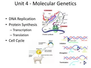
UNIT 3 Chapter 5: The Structure and Function of DNA Section 5.2 5.2 DNA Replication All life depends on the ability of cells to reproduce. Successful reproduction includes the transfer of the parent’s DNA to offspring. The life cycle of a cell is referred to as the cell cycle. For most cells, the cell cycle can be represented as in the diagram shown here. The process of copying one DNA molecule into two identical molecules is called DNA replication. DNA is copied during S phase of interphase in the cell cycle.
UNIT 3 Chapter 5: The Structure and Function of DNA Section 5.2 DNA Replication In the mid-1950s, three competing models of DNA replication were proposed: • conservative model • semi-conservative model • dispersive model The conservative model results in one new molecule and conserves the old. The semi-conservative replication model results in two hybrid molecules of old and new strands. The dispersive model results in hybrid molecules with each strand being a mixture of old and new strands.
UNIT 3 Chapter 5: The Structure and Function of DNA Section 5.2 Meselson and Stahl Determine the Mechanism of DNA Replication Matthew Meselson and Franklin Stahl reasoned that they could test the models if they could distinguish between parent and daughter strands of DNA. They used two different isotopes of nitrogen to label the DNA: “light” 14N and “heavy” 15N. These isotopes were chosen for two reasons: • Nitrogen is a component of DNA and would be incorporated into newly synthesized daughter strands. • “Light” and “heavy” forms of nitrogen would allow separation of different strands of DNA based on how much isotope was present. DNA with the “heavy” nitrogen would be more dense than DNA with “light” nitrogen. Continued…
UNIT 3 Chapter 5: The Structure and Function of DNA Section 5.2 Meselson and Stahl Determine the Mechanism of DNA Replication
UNIT 3 Chapter 5: The Structure and Function of DNA Section 5.2 DNA Replication Phase 1: Initiation Semi-conservative replication has three phases: initiation, elongation, and termination. In initiation, a portion of the DNA is unwound to expose the bases for base pairing. Unwinding starts at a specific nucleotide sequence called the origin of replication. Circular prokaryotic DNA has a single origin of replication while linear eukaryotic DNA often has thousands. Continued…
UNIT 3 Chapter 5: The Structure and Function of DNA Section 5.2 Initiation • Initiator proteins bind to the DNA and begin the process of unwinding. • Helicase enzymes cleave the hydrogen bonds that link the complementary base pairs. • Single-strand-binding proteins help to stabilize the unwound strands. • Topoisomerase II (in prokaryotes) relieves strain on the double helix that is generated from unwinding. A replication bubble forms with a Y-shaped replication fork at each end.
UNIT 3 Chapter 5: The Structure and Function of DNA Section 5.2 Elongation: Part I Two new strands are assembled using the parent DNA as a template. DNA polymerase III catalyzes the addition of new nucleotides to create a complementary strand to the parent strand. However, it can only attach new nucleotides to the free 3′ hydroxyl end of a pre-existing chain of nucleotides. Only one parent strand has a free 3′ hydroxyl end. The strand that is synthesized continuously in the 5′ to 3′ direction from this parent strand is called the leading strand. Continued…
UNIT 3 Chapter 5: The Structure and Function of DNA Section 5.2 Elongation: Part I
UNIT 3 Chapter 5: The Structure and Function of DNA Section 5.2 Elongation: Part II The lagging strand is formed in short segments away from the replication fork in a discontinuous manner. This requires primase to synthesize an RNA primer. Once the primer is attached to the parental strand, DNA polymerase III extends the strand by synthesizing DNA fragments called Okazaki fragments. DNA polymerase I removes the primers and fills in the space by extending the neighbouring DNA fragment. DNA ligase then joins the Okazaki fragments to create a complete strand.
UNIT 3 Chapter 5: The Structure and Function of DNA Section 5.2 Termination Termination occurs once the synthesis of the new DNA strands is complete. The two new DNA molecules separate from each other, and the replication machine is dismantled. At each replication fork lies a complex of proteins and DNA that make up the replication machinery. The image shown here is a simplified version of the molecules involved.
UNIT 3 Chapter 5: The Structure and Function of DNA Section 5.2 Important Enzymes in DNA Replication
UNIT 3 Chapter 5: The Structure and Function of DNA Section 5.2 Errors During DNA Replication A human cell can copy its entire DNA in a few hours, with an error rate of about one per 1 billion nucleotide pairs. Errors naturally occur during replication. Mispairing of bases and strand slippage are two types of errors that cause either additions or omissions of nucleotides. Incorrect pairing (mispairing) of bases is thought to occur as a result of flexibility in DNA structure. Continued…
UNIT 3 Chapter 5: The Structure and Function of DNA Section 5.2 Errors During DNA Replication Strand slippage during DNA replication can cause the addition or omission of nucleotides in newly synthesized strands, which represent errors.
UNIT 3 Chapter 5: The Structure and Function of DNA Section 5.2 Correcting Errors During DNA Replication DNA polymerase I and II have proofreading abilities. These enzymes recognize and correct errors in newly synthesized strands of DNA. This method repairs about 99% of the mismatch errors that occur during replication. Mismatch repair is done by a group of proteins that can recognize and repair deformities in newly synthesized DNA that feature the mispairing of bases. Errors that remain after DNA polymerase proofreading or mismatch repair are considered mutations once cell division occurs.
UNIT 3 Chapter 5: The Structure and Function of DNA Section 5.2 Comparing DNA Replication in Eukaryotes and Prokaryotes
UNIT 3 Chapter 5: The Structure and Function of DNA Section 5.2 Section 5.2 Review
UNIT 3 Biology Connections STSE Biobanking is the collection and analysis of physical specimens from which DNA can be derived from a wide sampling of individuals. A goal of biobanking is to provide researchers with open access to millions of high-quality tissue samples collected from people around the world. While biobanks offer the potential for gaining insight into the interaction between genes, environmental factors, and disease, they also present difficult legal and ethical challenges.