Wrist Injuries
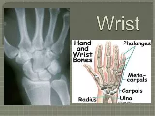
Wrist Injuries. Tintinalli Chapter 269. Wrist Anatomy. Wrist Radiography. Standard views: PA PA: 3 smooth arcs 2 arcs formed by proximal and distal surface of scaphoid, lunate, and triquitrum 3 rd arc: prox articular surface of capitate & hamate

Wrist Injuries
E N D
Presentation Transcript
Wrist Injuries Tintinalli Chapter 269
Wrist Radiography • Standard views: PA • PA: 3 smooth arcs • 2 arcs formed by proximal and distal surface of scaphoid, lunate, and triquitrum • 3rd arc: prox articular surface of capitate & hamate • Any distortion implies fx, dislocation, subluxation • Separation > 1-2mm b/w carpal bones indicates ligament disruption • Scaphoid shortening = ligament distruption or fx
Wrist Radiography • Standard Views • Lateral: 3 C’s • Axis of radius, lunate, capitate are collinear • Simple assessment of wrist dislocation, carpal allignment, degree of fx angulation • Oblique: partial pronation or supination • Scaphoid view
Ligamentous Injuries • Scapholunate Dislocation: most common • MOI: FOOSH impact on thenar eminence • pain with wrist hyperextension snap with radial or ulnar deviation, tender dorsum of wrist • Xray: PA • scaphoid has a dense ring “signet ring” • >3mm widening between lunate and scaphoid “Terry Thomas sign” • Tx: • Radial gutter splint • Ortho referral
Ligamentous Injuries • Lunate/Perilunate dislocation • MOI: FOOSH; requires greater force (MVA, fall from height) • Generalized swelling, pain, tender. Gross deformity absent • XRAY (key to dx) • Lateral: 3 C’s disrupted (spilled teacup sign) • PA: “piece of pie”sign • Emergency Ortho consult • Reducible: long arm splint • Irreducible: ORIF and ligament repair
Carpal Bone Fractures Scaphoid-most common • MOI • FOOSH or axial load along thumb metacarpal • Clinical • snuff box tenderness, pain with compression thumb metacarpal • XRay: standard and scaphoid view • Distortion of fat stripe • Tx • Nondisplaced-short arm thumb spica and ortho f/u • Displaced-Ortho for ORIF • Avascular necrosis possibility
Carpal Bone Fractures • Triquetrum-2nd most common • MOI: FOOSH or direct blow • Clinical: tender distal to ulnar styloid on the dorsal wrist • Tx-volar splint, ortho f/u • Lunate-3rd most common • FOOSH • pain middorsum of the wrist worse with axial compression of 3rd metacarpal • Tx-thumb spica, ortho f/u • avascular necrosis is a complication
Carpal Bone Fractures • Trapezium • MOI: direct blow to thumb • Clinically: painful thumb movement, weak pinch • Tx: • Nondisplaced thumb spica splint • Displaced >1-2mm ORIF • Pisiform • MOI: fall on hypothenar eminence • Clinically: tenderness on pisiform grasped b/w examiners fingers • Xray: carpal tunnel view; ossifies after age 12 • Tx: compression dressing or splint 30deg flexion and ulnar deviation
Carpal Bone Fractures • Hamate • MOI: interrupted swing of bat or golf club, handle impacts vs hypothenar eminence • Clinically: tenderness soft tissue of hypothenar eminence • Capitate (scaphoid fx in conjunction) • MOI: forceful dorsiflexion with impact on radial side • Clinically: swelling/pain proximal to 3rd metacarpal • Tx for above: • Nondisplaced: compression dressing or splint • Displaced: Ortho for ORIF • Trapezoid (rare)
Distal Radius and Ulna Fractures Colles Fracture • MOI: FOOSH • Xray: • Distal radial metaphysis fx • Distal fragment: displaced proximally and dorsally • Dinner fork deformity (lateral view) • May have Ulnar styloid fx • Tx: • Stable: splint, ortho closed reduction • Unstable: >20 degrees angulation, ortho for surgery
Distal Radius and Ulnar Fractures Smith’s fracture • MOI: direct blow dorsum hand/wrist, FOOSH hand in supination • Clinically: “garden spade deformity” • Xray: • Transverse fracture of the distal radial metaphysis • volar displacement of distal radius fragment • Tx: same as Colles fx