Download
1 / 34
380 likes | 1.76k Views
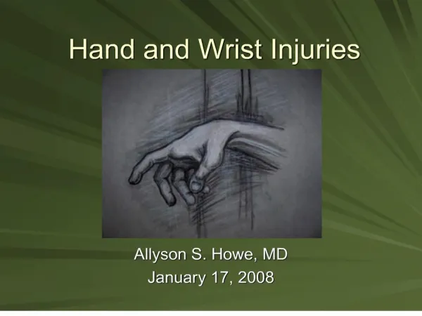
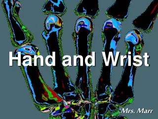
Chapter 12-Wrist and Hand Injuries. Objectives. Understand the basic anatomy of the wrist and hand. Explain various types of injuries that occur to the wrist and hand. Understand common mechanisms that cause injuries to the wrist and hand.

E N D
Objectives • Understand the basic anatomy of the wrist and hand. • Explain various types of injuries that occur to the wrist and hand. • Understand common mechanisms that cause injuries to the wrist and hand. • Understand the signs and symptoms of the various types of fractures of the wrist and hand.
Anatomy-Carpal Bones • “Some lovers try positions that they can’t handle” • Scaphoid, Lunate, Triquitral, Pisiform, Trapezium, Trapezoid, Capitate, Hamate • http://www.gwc.maricopa.edu/class/bio201/hand/awrist.htm
Anatomy-metacarpals and phalanges • Labels 1-5, beginning at thumb • 5 metacarpals (each hand) • 14 phalanges (each hand) • Phalanges classified as proximal, middle and distal phalanx
Anatomy-joints • Named for the bones that comprise them • Carpometacarpal (CMC) • Metacarpal phalangeal (MCP) • Proximal interphanlageal (PIP) • Distal interphalangeal (DIP) • http://www.gwc.maricopa.edu/class/bio201/hand/anhand.htm
Anatomy-muscles • flexor groups
Anatomy-muscles • Extensors
Anatomy-hand • Adductors and Abductors of phalanges
Anatomy-muscles • Thenar eminence
Anatomy-muscles • Hypothenar eminence
Anatomy-Ligaments • Mostly intrinsic due to need for bones to be connected • Radial and ulnar collateral • Flexor retinaculum or transverse carpal ligament • Stablizes carpals, covers flexor tendons and median nerve
Anatomy-ligaments • Thumb • Ulnar collateral of the thumb • Radial collateral of the thumb • Provide joint stability to thumb
Injuries • Prevention • Braces • Tape • Gloves • Padding
Ligament Injuries • Wrist sprains • Often caused by overuse, falls, and forceful twisting motion • Injury side depends on side of twist/overstretch • S/S- pain, decreased ROM, decreased grip strength, possible swelling • PRICE • Taping for support • Rehab focuses on establishing normal ROM and return to normal strength
Dislocated lunate • Most commonly dislocated carpal bone • Causes deformity, pain, swelling, and decreased ROM • Splint and refer • Physician relocates
Ganglion Cyst • Fluid within the muscle sheath • Treat with ice and activity modification • Refer to physician if symptoms persist
Gamekeeper’s Thumb • Injury to the medial collateral ligament • aka skier’s thumb • Usually caused by forced abduction • Pain and swelling likely • Treat with ice and splinting of medial aspect of thumb • Should have x-ray
Interphalangeal collateral ligament sprains • “jammed” finger • Often caused by finger hit by ball • Swelling, pain, discoloration • May refer for x-ray • Ice, buddy tape, possible padding
Dislocations of ICP or MCP joints • One bone usually moves volar (palm side), and one dorsal • Refer to physician for relocation because of tiny tendons, nerves and blood vessel movement • Improper relocation can cause permanent damage
Tendinitis • Inflammation of tendon • Caused by overuse, stretching or impact • Prevent by increasing strength and flexibility
De Quervain’s tendinitis • Affects abductor pollicis longus and extensor pollicis brevis • Shot-putters are vulnerable • Difficulty abducting, swelling, crepitis • Limit activity, PRICE
Mallet finger • Result of direct impact • Caused by tearing of extensor tendon from bone • Cannot extend tip of finger due to tendon disruption • Pain, swelling, need to splint in extension and refer • Possible treatment- sugery or splint • If not treated, finger will remain in permanent flexion
Jersey finger • Similar to mallet finger except injury it to flexor tendon • Inability to flex DIP joint • Named from moi, holding jersey with fist and finger forced to extend, tearing tendon • Causes pain and swelling • Splint, ice, and refer
Boutonniere deformity • Occurs at PIP join • Caused by hard impact over PIP, results in tear in the joint capsule, extensor tendons fall laterally • Extensors contract and force flexion of the PIP and extension of DIP • Pain and swelling • Splint and refer • Continued splint or surgery are treatments
Scaphoid fracture • Commonly caused by falling on outstretched hand • Pain, swelling, decreased ROM • Refer for x-ray • Casting common • Untreated or improperly treated can cause a non-union fracture and/or avascular neucrosis
Complications of scaphoid fracture • Avascular neucrosis • lower half of the fractured bone loses its blood supply and actually dies • Can eventually lead to degenerative arthritis • Non-union fracture • If fx does not heal properly, surgery may be required
Boxer’s fracture • most common type of metacarpal fracture • point of maximal tenderness is just proximal to the knuckle • Caused by direct blow/punch • Ice, splint, and refer
Colle’s fracture • caused by falling forward onto an outstretched arm • sometimes called a 'dinner fork‘ • Splint and refer for x-ray and treatment
Smith’s fracture • Similar to Colle’s fracture, but wrist in flexion • patient lands with the wrist in flexion
Carpal tunnel syndrome • caused when too much pressure is put on the median nerve • Check with Tinel’s test or Phalen’s test • Refer to physician for recommended treatment