Download

1 / 33
330 likes | 576 Views
Educational Initiatives in AMD and Related Retinal Diseases. What is AMD?. AMD is a degenerative retinal disease that can cause central vision loss AMD is the leading cause of severe vision loss in people over 50 years of age. Lens. Retina. Optic nerve. Macula. AMD Damages the Macula.

E N D
What is AMD? • AMD is a degenerative retinal disease that can cause central vision loss • AMD is the leading cause of severe vision loss in people over 50 years of age
Lens Retina Optic nerve Macula AMD Damages the Macula
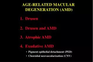
Two Forms of AMD Can Cause Severe Vision Loss Non-neovascular (geographic atrophy) Neovascular (choroidalneovascularization)
Progression of Neovascular AMD:Normal Retina Photoreceptors RPE Choroid
Progression of Neovascular AMD: Development of Drusen Bruch’s membrane thickens and drusen develop
Progression of Neovascular AMD:Formation of New Vessels New abnormal blood vessels proliferateand penetrate Bruch’s membrane
Progression of Neovascular AMD:Leakage of Fluid and Blood from CNV New blood vessels leak blood and fluid
Symptoms of AMD • Reduced central vision • Central scotoma • Distortion • Decreased contrast sensitivity • Decreased color vision
Impact of AMD on the Patient:Visual Function Patients with neovascular AMD may have difficulty with visual tasks: • Reading • Telling the time • Recognizing faces • Driving
Public Awareness of AMD Despite the prevalence of AMD and its impact: • Only 4% of adults are ‘very familiar’ with AMD • Only 2% of adults know that AMD is the leading cause of vision loss in people over 50 years of age • 75% of people do not know of a treatment for AMD • Almost 40% of people have eye examinations less often than every 2 years
Management of AMD • Early detection and treatment are important • Proven treatments are available for many patients with neovascular AMD • Standard (thermal) laser photocoagulation • Verteporfin (Visudyne™) therapy • Patients who experience vision loss may benefit from low vision aids and education
Verteporfin Therapy:A Two-Step Process Step 1 Step 2
Outcomes in Clinical Trials of Verteporfin Therapy • Safely reduces risk of moderate and severe vision loss • Most patients lose some vision over 2 years after initiating therapy due to natural course of disease • Modest improvements in vision can occur, but are unusual • Verteporfin therapy should be considered in selected patients, depending on fluorescein angiogram • Retreatment is required in most patients, as often as every 3 months (average 5–6 treatments over 2 years)
Patient Selection for Verteporfin Therapy To identify who may benefit from verteporfin therapy, diagnostic assessment should include: • Best-corrected visual acuity • Fundus biomicroscopy via a dilated pupil • Color fundus photography • Fluorescein angiography
Preparing the Patient Patient instructions should provide information about: • Two-step procedure • Measuring height and weight • Realistic expectations • Photosensitivity precautions • Follow-up and retreatment
Verteporfin Infusion Kit • Sterile water • D5W • Tubing • Filters • Needles • Catheter • Syringes • Alcohol wipes
Verteporfin Formulation Verteporfin is a light-activated drug: • Supplied in single-use 15 mg vials • Sterile, lypophilized, dark-green cake • Stored at room temperature(20–25ºC or 68–77ºF)
Verteporfin Reconstitution Verteporfin is reconstituted with sterile water for injection • 7 mL of water is added to the vial to give a volume of 7.5 mL • The vial is gently agitated to ensure complete dissolution • The solution must be used within 4 hours • Concentration of reconstituted drug 2 mg/mL
Calculating BSA Height Weight BSA (e.g.: 1.88 m2)
Calculating the Verteporfin Dose Drugdose BSA 6 mg/m2 e.g. 1.88 m2 Totaldrugdose Reconstituteddrugconcentration Volume reconstituteddrug = e.g. 11.28 mg 2 mg/mL e.g. 5.64 mL
Preparing the Infusion • The appropriate volume of verteporfin solution is withdrawn from the vial • The verteporfin is transferred to the 30 mL syringe • D5W is added to give the final 30 mL volume for infusion
Infusion Connections Syringe in pump • A filter is fitted to the syringe • The syringe is connectedto the IV line Infusion tubing Venous access Filter Needle
Establishing the IV Line A free-flowing IV line is established, preferably in the antecubital vein
Infusing the Verteporfin Solution • An infusion or syringe pump delivers the 30 mL of drug over 10 minutes • The timer on the laser system is started at the same time as the infusion
Precautions to Avoid Extravasation • Extravasation may cause severe pain, inflammation, swelling, and discoloration of the injection site • The IV line should be carefully monitored during treatment and infusion stopped if extravasation is suspected or recognized • Even with good technique, extravasation may occur
Procedure in the Event of Extravasation • If more than half the dose delivered • Proceed with light application • If less than half the dose delivered • Obtain better venous access • Begin light application 15 min after restarting infusion • Apply cold compress or ice immediately • Protect from light for at least 5 days or as long as skin is discolored • Consult burn specialist or dermatologist if needed
Ending the Infusion • The pump is turned off • The IV line is flushed with 5 mL D5W until all remaining verteporfin is cleared from the line • The laser timer will indicate when to begin light application
Positioning the Patient and Applying the Light • The patient is positioned at the slit lamp immediately after the end of infusion Light at 689 nm is delivered via a fiber optic and slit lamp using a contact lens Light dose • 600 mW/cm2 • 83 seconds 50 J/cm2
Photosensitivity Precautions Patients are advised to: • Avoid exposing unprotected skin to direct sunlight or bright indoor light according to physician’s instructions • Wear dark sunglasses • Wear wide-brimmed hat, long sleeves, and pants while outdoors • Reschedule elective or dental surgery after treatment • Wear wristband provided, to indicate potential photosensitivity in event of emergency surgery
Patient Education Physician needs to manage patient expectations: • Treatment is designed to reduce the risk of vision loss • Vision improves in few cases • Retreatment is often required at 3-month intervals during the first 2 years
Summary • Staff members have an important role in helping to ensure that treatment is administered correctly • Staff members reinforce information to ensure that patients: • Understand the treatment process • Follow the precautions • Have realistic expectations of treatment outcomes
This activity was made possible by an unrestricted educational grant from Novartis Ophthalmics, Inc.