Download
1 / 18
190 likes | 881 Views
AGE-RELATED MACULAR DEGENERATION (AMD). 1. Drusen. 2. Drusen and AMD. 3. Atrophic AMD. 4. Exudative AMD. Pigment epithelial detachment (PED). Choroidal neovascularization (CNV). Drusen. Histopathology. Hard. Soft. Larger, ill-defined spots. Small well-defined spots.

E N D
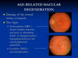
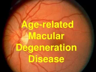
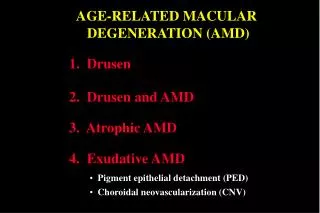
AGE-RELATED MACULAR DEGENERATION (AMD) 1. Drusen 2. Drusen and AMD 3. Atrophic AMD 4. Exudative AMD • Pigment epithelial detachment (PED) • Choroidal neovascularization (CNV)
Drusen Histopathology Hard Soft • Larger, ill-defined spots • Small well-defined • spots • May enlarge and coalesce • Usually innocuous • Increased risk of AMD
`` FA of drusen Degree of hyperfluorescence depends on: • Extent of overlying RPE atrophy (window defect) • Amount of staining • Lipid content
Drusen and AMD - progression Exudative AMD Atrophic AMD
Atrophic AMD Progression Initially drusen and non-specific RPE changes Late RPE (geographic) atrophy
Atrophic AMD Fluorescein angiogram Management Hyperfluorescence from RPE window defect Low-vision aids if appropriate
Signs of Pigment epithelial detachment Sub-RPE fluid may be clear or turbid Circumscribed, dome-shaped elevation
FA of pigment epithelial detachment No increase in size of lesion Progressive increase in hyperfluorescence Early, well-defined hyperfluorescence
ICG angiogram of pigment epithelial detachment Later, thin surrounding hyperfluorescent ring Early, well-defined hypofluorescence No increase in size of lesion
Possible subsequent course of PED Spontaneous resolution Geographic atrophy CNV RPE rip
Choroidal neovascularization (CNV) • Less common than atrophic AMD but more serious • Metamorphopsia is initial symptom • Most lesions are not visible clinically Suspicious clinical signs Subretinal blood or lipid Pinkish-yellow subretinal lesion with fluid
Angiographic classification of CNV Well-defined (classical) Occult • Extrafoveal > 200 m from centre of • FAZ • Poorly defined • Juxtafoveal < 200 m from centre of • FAZ • Obscured by PED, blood or exudate • Subfoveal - involving centre of FAZ
FA of classical CNV Leakage into subretinal space and around CNV Late staining Very early ‘lacy’ filling pattern
ICG angiogram in PED with occult CNV PED is hypofluorescent CNV is hyperfluorescent (hot spot)
Possible subsequent course of CNV Subretinal (disciform) scarring Haemorrhagic sensory and RPE detachment Massive subretinal exudation Exudative retinal detachment
Potential indications for laser treatment of CNV • Classic extrafoveal CNV on FA • Occult extrafoveal CNV on ICG Pre-treatment FA of classic CNV
Technique of laser photocoagulation of CNV • Perimeter is treated with overlapping 200 m (0.2-0.5 sec) burns • Entire area is covered with high energy burns Late staining around margin is normal Lack of leakage following successful treatment
Results of laser photocoagulation of CNV • Initial risk of severe visual loss reduced by over 50% • Frequent subsequent recurrence with subfoveal involvement Recurrence of CNV several months after initially successful treatment