Vision
Vision. Dr. Janet Fitzakerley jfitzake@d.umn.edu http://www.d.umn.edu/~jfitzake/Lectures/Teaching.html. OPTICS RETINA PHYSIOLOGY. Which of the following is MOST LIKELY to occur in Parkinson disease? One best answer. Decreased corneal reflex due to death of neurons in the trigeminal ganglion


Vision
E N D
Presentation Transcript
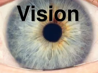
Vision Dr. Janet Fitzakerleyjfitzake@d.umn.eduhttp://www.d.umn.edu/~jfitzake/Lectures/Teaching.html
Which of the following is MOST LIKELY to occur in Parkinson disease?One best answer • Decreased corneal reflex due to death of neurons in the trigeminal ganglion • Decreased corneal reflex due to macular degeneration • Decreased blinking due to loss of dopaminergic input to premotor brainstem structures • Decreased sympathetic drive from the superior cervical ganglion • Increased acetylcholine release fromfacial and oculomotor neuronsinnervating OO and LPS
The cornea and lens focus light on the retina; the cornea has greater refractive power but the focusing power of the lens can be adjusted to allow near vision (accommodation). Refractive errors include cataracts, hyperopia, myopia, presbyopia and astigmatism. REFRACTION
Which of the following involves the image being focused inappropriately in front of the retina?More than one “correct” answer – but one best answer! • Astigmatism • Cataracts • Emmetropia • Hyperopia • Myopia • Presbyopia
How would you correct the problemin the previous question?One best answer! • Contact lenses that correct refraction inone meridian • Glasses with positive diopters • Refractive surgery to flatten the cornea • Replacement of lens • Reading glasses • No need to do anything
Light intensity is regulated by the PUPILLARY LIGHT REFLEX, which causes MIOSIS as a result of parasympathetic stimulation of the sphincter pupillae muscles (muscarinic receptors). MYDRIASIS results from sympathetic stimulation (α1 receptors) that activates the dilator pupillae muscles. PUPILLARY LIGHT REFLEX
Where is the damage if you shine a bright lightinto a person’s left eye,and the right pupil DOES NOT constrict?More than one “correct” answer • Photoreceptors in the left eye • Photoreceptors in the right eye • Left optic nerve • Right optic nerve • Left optic tract • Right optic tract • Left visual cortex • Right visual cortex • Left oculomotor nucleus • Right oculomotor nucleus • Left dilator pupillae muscle • Right dilator pupillae muscle • Left sphincter pupillae muscle • Right sphincter pupillae muscle
http://www.youtube.com/watch?v=dotS3BaEH8s If I told you that the person’s left eye constricts in responseto light in the left eye, would that help you localize the lesion?
Which of the following would result in increased intraocular pressure?One “correct” answer • Decreased carbonic anhydrase activity • Increased parasympathetic activity • Relaxation of the ciliary muscle • Stimulation of α1 receptors • Stimulation of β2 receptors
Increased intraocular pressure causes loss of vision (potentially permanent). Open angle glaucoma (the most common form) results from overproduction of the aqueous humor. Closed angle glaucoma (typically the most rapidly evolving form) is caused by blockage of fluid outflow.
Which of the following isMORE CHARACTERISTIC of rodscompared to cones?One “correct” answer • Detection of far red wavelengths • Location in the fovea • Only activated by multiple photons • Fast responses • Response to scattered light • Lack of convergence (in terms of output from retina)
Astronomers know that the best way to see faint nebulae and star clusters is to avert their vision (at night, you can see a dim object better if you look a bit to one side)? Which of the following BEST explains this phenomenon?One “correct” answer • Rods have better spectral sensitivity than cones. • Rods have better temporal sensitivity than cones. • Rods have better spatial acuity than cones. • Rods undergo dark adaptation more than cones.
The ability to discriminate fine details of the visual scene is termed VISUAL ACUITY. Three types are recognized: SPATIAL, TEMPORAL and SPECTRAL. Visual acuity is primarily a function of the cone system. Rods are responsible for SCOTOPIC vision (the monochromatic vision that occurs in low light). The three types of cones (blue, green and red; or Short, Medium and Long wavelength) have better temporal and spatial resolution than rods, making PHOTOPIC VISION better for discrimination of surfaces and movement under bright light conditions.
How is transduction in olfactory receptor neurons similar to rod phototransduction? Both systems:One “correct” answer • have multiple receptor proteins in individual receptor cell. • use the same G-protein. • activate phosphodiesterase. • involve ligand-gated non-selective cation channels. • result in depolarization following receptor activation.
PHOTOTRANSDUCTION occurs via a 4 step process that uses a 2nd messenger cascade to amplify the signal. In rods, activation of rhodopsin ultimately results in the closureof cyclic nucleotide gated Na+ channels, and hyperpolarization of the photoreceptor.
Which of the following occurswhen low intensity light hits the photoreceptors?One “correct” answer • Rhodopsin is cleaved but cone pigments are not. • An opsin and all-trans-retinal are formed from rhodopsin. • The retinal pigment epithelium takes up rhodopsin. • Rods accumulate rhodopsin. • Rhodopsin is reformed in the retinal pigment epithelium from an opsin and all-trans-retinal.
The VISUAL CYCLE consists of bleaching and recycling of 11-cis-retinol between the photoreceptors and the retinal pigment epithelium (RPE). It is a key component of dark adaptation in rods and is disrupted in vitamin A deficiencyand macular degeneration.