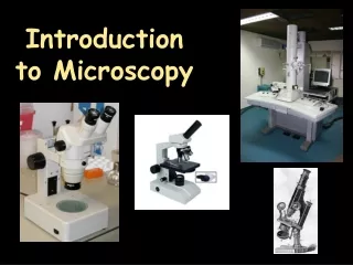
Introduction to Microscopy
Introduction to Microscopy. Types of Microscopes. Dissection or Stereoscope SEM ( Scanning Electron Microscope) TEM ( Transmission Electron Microscope) Compound ( Light) Microscope***. Difference between Magnification and Resolution.


Introduction to Microscopy
E N D
Presentation Transcript
Introduction to Microscopy
Types of Microscopes • Dissection or Stereoscope • SEM ( Scanning Electron Microscope) • TEM ( Transmission Electron Microscope) • Compound ( Light) Microscope***
Difference between Magnification and Resolution • Magnification: is just power of an instrument to enlarge things...Just as you would increase the size of a picture in your PC by zoom function. It would enlarge it... • Resolution : is power to show two closely situated particles as different... that obviously improves quality of the picture.. • The magnification of human eye is 1 and the resolution is 1 micrometer.i.e. The eye does not magnify a thing and two particles spaced at a distance of 1 micrometer will be able to be seen different.No wonder objects closer than this will be seen as one. • Human hair: 50 micrometers
Dissection or Stereoscope • Used to get a better look at a larger specimen • Low magnification- 10X- 40X • 3-D image • Light illuminated
Scanning Electron MicroscopeSEM • Used to examine very small, multicellular, anatomical structures • 3-D structures • Specimen is coated with metal • Electron beam is swept across specimen causing electrons from specimen to get excited causing secondary electrons to be emitted and collected • Electrons are detected and an image is created • High Magnification/High Resolution
Scanning Electron Microscope Follicle cell to nurture cell Human Egg Sitting on the point of a pin-
Scanning Electron Microscope Magnification: X 500 Black Widow Spider Claw
Scanning Electron Microscope Mascara brush with mascara and flakes of skin-50X
Scanning Electron Microscope Velcro- hook and loop-35X
Transmission Electron Microscope • Works similar to a slide projector: beam of electrons ( not light) passes through specimen and projected on screen below. • 2-D view of internal organelles • Specimen sliced thin • Very high resolution and high magnification. Much higher than a compound microscope
Transmission Electron Microscope Normal Collagen Degenerating Collagen A pair of images of collagen fibrils in the eye of a patient with glaucoma revealed.
Transmission Electron Microscope Cell undergoing Apoptosis- committing suicide
Compound Light Microscope • Light Illuminated • Image is 2-D • High Magnification but low resolution • View cells that are living- example algae • Tissue samples may be sectioned, stained and put onto a slide • There are several different magnification lenses
Compound Light Microscope Frog’s blood- 400X Magnified
Compound Light Microscope Frog’s Blood- 1000X Magnified
high=40X; low=10X; scanning=4X changes objective lenses directs light into microscope focuses light amount of light entering the microscope brings object into slow focus look through; lens is 10X carry and support supports slide; positions slide brings object into rapid focus
Type of Light Microscopy • IMMUNOFLUORESCENCE • Specific molecules are located within the cell by an antibody produced against the molecule of interest. • The antibody- fluor attaches to the molecule and under a light microscope the molecule can be observed. • This can be used in order to see whether certain molecules are present or not present and at what concentration.