Download
1 / 52
930 likes | 2.13k Views
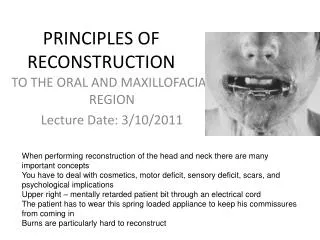
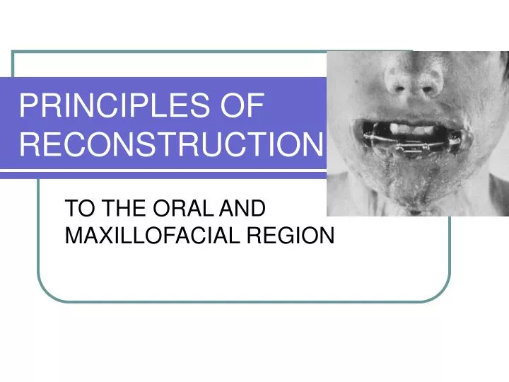
PRINCIPLES OF RECONSTRUCTION. TO THE ORAL AND MAXILLOFACIAL REGION. RECONSTRUCTIVE CHALLENGE. DEFECTS OF THE FACIAL BONES. TYPES (5) 1. Congenital 2. Pathologic 3. Trauma related 4. Infectious 5. Acquired. Each defect poses its own unique challenges.

E N D
PRINCIPLES OF RECONSTRUCTION TO THE ORAL AND MAXILLOFACIAL REGION
DEFECTS OF THE FACIAL BONES • TYPES (5) 1. Congenital 2. Pathologic 3. Trauma related 4. Infectious 5. Acquired
MOST MAXILLOFACIAL DEFECTS Will have both an osseous and soft tissue component
OSSEOUS RECONSTRUCTION • Functions of the mandible: a) Facial contour b) Facial support c) Muscles of mastication d) Tongue/Floor of mouth e) Muscles of facial expression
INTRODUCTION TO THE CANCER RELATED DEFECT • NEEDS: Bone Soft Tissue Augmentation of existing ridges Removal of scar and mucosal encombrances Restoration of facial form
BONE GRAFTS • Healing of bone is unique: new bone arises from tissue regeneration with cellular proliferation (osteoblasts) and collagen synthesis
BONE HEALING CONCEPT Vascularization 2 Phases of Healing Axhausen 1956
PHASES • Phase 1: starts from transplanted cells in the graft that proliferate and form new osteoid. Determines the quantity of surviving bone • Phase 2: consists of the classical resorptive remodeling process- no new quantitive bone is formed
Healing overview Superficial cellular elements survive by nutrient diffusion Hematoma formation Granulation tissue ingrowth Prominent vascular and mesenchymal cellular components ie OSTEOINDUCTION
Healing con’t • Osteoclastic activity allows further vascular invasion. • Osteoinduction occurs:mesenchymal tissue differentiates into tissue capable of osteogenesis-regulated by BMP • Osteogenesis with repair and incor- poration of graft = final phase
ACTIVATION OF BONE REPAIR • Occurs by local and systemic factors Parathyroid hormone: most potent agent to stimulate remodeling activity. Stimulates pluripotential cells (elaborates osteoclasts). Ca++, GH,thyroxin stim. activity. Calcitonin,estrogen, adrenocortical steroids inhibit activity
BONE FORMATION Stimulated by growth factors
Osteogenin BMP-3 BMP-2A BMP-2B BMP-5 BMP-6 Osteogenic protein BMP-7 TGF-B1-5 Vg-1Activin Beta A & B Inhibin alpha Decapentaplegic complex Mullerian inhibiting substance TRANSFORMING GROWTH FACTOR-BETA SUPERFAMILY
TGF-B SUPERFAMILY • Overall Function: osteogenesis, chondrogenesis, wound healing, fx healing, cellular differentiation ie multifunctional growth factor
TYPES OF GRAFTS • Vascularized ie osteomyocutaneous • Nonvascularized cortical cancellous other:bone slurry,particulate bone bone paste(paté) corticocancellous
CLASSIFICATION BY STRUCTURE • Cortical • Cancellous • Corticocancellous • Cortical vs cancellous re: ease of vascular penetration
CLASSIFICATION BY ORIGIN • Autogenous/Autograft • Allograft/Isograft/Homograft:one individual to another of the same species. Lyophilized—reconstitute • Xenograft:tissue transplant from 1 species to another. Bio-Oss/Osteogen • Alloplasts:Inert synthetic materials
ALLOGRAFTS • Are osteoconductive (no viable cells) • Packaged in a lyophilized state and must be reconstituted • exam: freeze dried, demineralized bone • Offers only a hard tissue matrix for the inductive phase of healing (no viable cells for osteogenesis)
SOFT TISSUE BED • Is the key to reconstruction of head and neck patients • Fibroblastic cellularity and vascular density determine the quality of the recipient bed • Concept of ORN and HBO
State of the Art: Bone grafts? • Primary cancer resection with post-op RT • Wait 1 year • Hyperbaric oxygen protocol with allogenic crib and iliac crest PCBM mandibular reconstruction • Wait 4-6 months • Vestibuloplasty and placement of osseointegrated implants • Wait 4-6 months • Prosthetic reconstruction Marx, RE. JOMS, 1995
Who Needs Bone? • Pre- free flap era • 46% success rate with non-vascularized grafts in primary mandible reconstruction • Success improved to 90% with delayed repair when graft placed extra-orally • Multiple surgeries required • Limited number of patients achieved functional reconstruction Lawson, W, et al. Laryngoscope 1982
TIMING OF RECONSTRUCTION • Two choices a)Delayed: wait a period of time post-ablation and then return patient to OR for reconstruction(usu. 6 months) Radiation, scarring, contracture,muscle atrophy, increased hospitalization,poor cosmesis and fibrosis can contribute to a suboptimal result
DELAYED RECONSTRUCTION • Due to masticatory muscle pull, typical distractive forces produce an upward and medial rotation of the bone segments • The primary advantage of delayed reconstruction is to avoid wound contamination by saliva
TIMING OF RECONSTRUCTION • b) immediate: primary reconstruction is best accomplished by free tissue transfer with less scarring, fibrosis, fewer hospitalizations, and decreased complication. Will see clear benefits with respect to cosmesis, oral competence, deglutition/mastication, speech, sense of wholeness and dental rehabilitation
IMMEDIATE RECONSTRUCTION • Immediate single-stage reconstruction is always preferable to delayed reconstruction • Prevents development of muscle contracture
ONCOLOGIC RECONSTRUCTION • 20,000 NEW CASES OF ORAL CAVITY CARCINOMA OCCURS ANNUALLY IN US • Primary mode of treatment is surgical, and 50% of oral cavity cancers are in the advanced stages
MICROVASCULAR FREE TISSUE TRANSFER • Can tailor the donor flap to the specific needs of the ablative defect • Not restricted by arc of rotation • With bone-containing flaps, resorption is minimized • Possibility of neural anastomosis • Only true contraindications is hypercoagulable states
ARTERIAL AND VENOUS ANASTOMOSIS • Arterial connections into external carotid artery system, most commonly the facial artery, and also the superior thyroid artery • Recipient veins include the internal and external jugular veins, facial veins or cephalic vein from arm
MANDIBULAR RECONSTRUCTION • Resection of mandible produces deformity and disability: facial contour/support; mm of mastication/facial expression,tongue/FOM support • Also, airway concerns: site of insertion of suprahyoid musculature which elevates the larynx and prevents aspiration; mand. supports tongue and thus the oral airway
MANDIBULAR RESECTION • Mandible can be resected: subtotal or marginal, continuity defect, anterior/lateral/angle/condyle-ramus unit • Primary free tissue transfer usually by fibula osteoseptocutaneous flap(peroneal vessels) or the iliac flap based on the deep circumflex iliac vessels • RIGID FIXATION MANDATED
Anterior Defect • No reconstruction • “Andy Gump” deformity • Loss of lip support • Loss of oral competence • Loss of function and cosmesis
ANDY GUMP DEFORMITY • Resection of the mandibular symphysis/ anterior mandibular arch • Oral competence suffers from the patient’s inability to manage oral secretions, speak,eat, or swallow • Most important deformity to reconstruct primarily
OTHER MANDIBULAR DEFORMITIES • Posterior-lateral mandible, angle, and the ascending ramus defects can be effectively treated with free tissue transfer or with conventional techniques
EXTIRPATIVE DEFECTS • Mandibulectomy defects • Maxillectomy defects • Glossectomy defects • Defects resulting from excision of oral lining tissues i.e. floor of mouth,pharyngeal wall and cheek
MAXILLARY RECONSTRUCTION • Try to achieve three (3) goals 1. Restoration of facial form-support for the orbital contents, cheek and lip 2. Separate sinus/nasal cavity from oral cavity 3. Dental rehabilitation
TONGUE RECONSTRUCTION • Most difficult of reconstructive challenges • Need to fulfill the requirements of: articulation mastication/swallowing laryngeal protection Must have a flap that is thin, mobile and hopefully can be made sensate.
SPEECH CONCERNS • Anterior tongue restriction: distorts “d” and “t” • Posterior tongue restriction: distorts “g” and “k” • Scar of lower lip distorts “v” and “f” • Components of speech are: respiration, phonation,resonance, articulation and neurologic integration